STORM - A new tool for scientists to study the physical changes underlying the development of long-lasting pain
Full Reference: Woodhams, Stephen G.a,*; Markus, Robertb; Gowler, Peter R.W.a; Self, Timothy J.b; Chapman, Victoriaa Cell type–specific super-resolution imaging reveals an increase in calcium-permeable AMPA receptors at spinal peptidergic terminals as an anatomical correlate of inflammatory pain, PAIN: November 2019 - Volume 160 - Issue 11 - p 2641-2650 The full article can view viewed online.
The development of long-lasting pain involves changes in the strength of connections between the nerve cells that conduct sensory information from the body to the brain. One of the key goals in pain research is to identify how and when these connections are altered, in order to find ways to prevent or reverse this process and thus provide pain relief. A key site where this occurs is the spinal cord, where incoming information from the nerve fibres in the body are received by spinal nerve cells to encode the location and strength of a painful stimulus, before sending it on to the brain. Damage to tissues of the body or the nerve fibres themselves increases the strength of these connections, meaning sensory stimuli such as light touch and moderate temperatures can now be perceived as painful (allodynia), and normally painful stimuli become even more painful (hyperalgesia) over time.
Activity in incoming nerves and their spinal partners can be assessed by recording the electrical currents during sensory stimulation, but this involves invasive procedures which may alter the connections between these cells, and also only allows for a few cells to be recorded at any one time. An alternative assessment method is to quantify changes in the number of key signalling molecules, such as receptors and ion channels, at the connections between cells (synapses), but this is difficult as the individual molecules are too small and close together to be assessed by conventional methods.
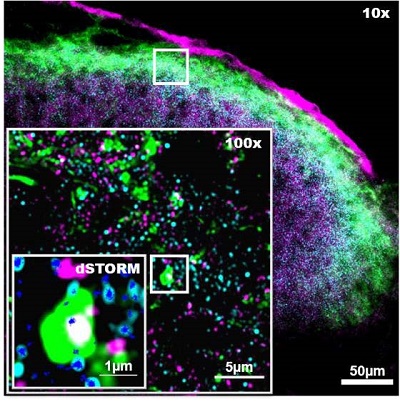
A recent study from Pain Centre Versus Arthritis at the University of Nottingham used a new kind of microscope with increased resolution (stochastic optical reconstruction microscopy; STORM) to look for changes in receptors in a mouse model of inflammation and pain.
STORM imaging revealed that pain behavior was accompanied by increased expression of pain-associated receptors in the spinal cord, a change that was not detectable byconventional microscopy alone. Looking at specific nerve cell types revealed that many of these receptors were associated with pain sensing nerve fibers. The amount of pain-associated receptors in these cells correlated with the degree of pain sensitivity in individuals, suggesting that this receptor can be used as a marker of increased synaptic strength and pain sensitivity.
This study was the first use of this kind of microscopy in pain research, and the first time it was applied to spinal cord tissue, adding a new tool for scientists to study the physical changes underlying the development of long lasting pain in the future.