In Vivo Microscopy of the Nervous System
In Vivo Microscopy (IVM) is a rapidly developing field in which real-time images of tissues taken in vivo provide cellular-level information, on the function and structure of cellular (e.g. neuronal) and non-cellular (e.g. vascular) networks.
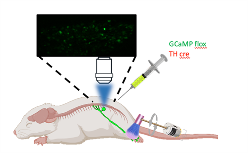
Pathophysiological changes in tissues, including changes to the peripheral and central nervous system, contribute substantially to the onset and maintenance of chronic pain. Changes in Dorsal Root Ganglia (DRG) are of importance because they contain the neurons that are responsible for the sensations of pain, touch, temperature, and proprioception. The spinal cord in turn takes this information from the DRGs and modifies and integrates it to send it on to the brain.
We have used in vivo microscopy to record large populations of neurons in the DRG and spinal cord, providing more information than possible with single-unit recordings. With the use of advanced genetically encoded calcium indicators large populations of DRG and spinal cord neurons can be assessed in response to a range of electrical, thermal and mechanical stimuli.
If you are interested to know more about In Vivo Microscopy used within Pain Centre Versus Arthritis, please contact Kim Chisholm.
View the publications that have used In Vivo Microscopy