Self-funded student vacancies
This is a list of research opportunities for international students or for UK students who can cover their own fees and living expenses. If you need information on scholarships, check our international scholarships page.
Find out how to apply
How to use the table
Clicking on a member of staff's name will take you to their personal home page whereas clicking on a PhD title will show more details about that particular project.
Research Opportunites (Self-funded students)
| Staff | Project title |
|---|
| Cameron Alexander |
Advanced responsive polymers for drug delivery |
| |
Synthetic polymer-cell interactions |
| Morgan Alexander, Amir Ghaemmaghami |
Promoting wound healing and re-epithelisation through surface engineering |
| Tracey Bradshaw |
Investigating antitumour activities and mechanisms of action of novel molecules isolated from Malaysian Rainforest flora. |
| Jonathan Burley |
Raman microscopy for characterising solid dosage forms |
| |
Extreme miniaturisation for discovering new formulations |
| Cynthia Bosquillon |
Understanding the fate of inhaled medicines in the lung |
| |
Evaluation of new biodegradable polymeric nanoparticles for drug delivery to the lung |
| Weng Chan |
Novel enabling chemical methods to macrocyclic peptides and their evaluation as antimicrobials |
| Cristina De Matteis |
New treatments for tuberculosis |
| Cornelia De Moor |
mRNA metabolism as a target for anti-inflammatory drugs |
| |
Finding the target of cordycepin in the PI3K/mTOR/AKT signal transduction pathway |
| Cornelia De Moor, Dong-Hyun Kim |
The function of secondary metabolites from insect infecting fungi |
| Cornelia De Moor, Keith Spriggs |
The function of polyadenylated long non-coding RNAs |
| Cornelia De Moor, Michael Stocks |
Developing novel polyadenylation inhibitors as lead compounds for the treatment of cancer and pain |
| Cornelia De Moor, Pavel Gershkovich |
Pharmacokinetics and pharmacodynamics of cordycepin, a potential lead drug in osteoarthritis |
| Lodewijk Dekker |
Overcoming treatment resistance in pancreatic cancer |
| James Dixon |
Development of Gene-activated, Injectable and 3D-prinatble bone repair matrices |
| |
Reprogramming Stem cells using GET peptide technology |
| |
Oral Insulin delivery using GET peptide technology |
| |
Light-inducible delivery using GET peptide technology |
| |
CRISPR/Cas9 gene-editing using GET peptide technology |
| Ingrid Dreveny |
Structure-based approaches for ubiquitin specific protease functional analysis and drug discovery |
| |
Proteases in the ubiquitin system - from molecular mechanisms to the development of novel inhibitors |
| Jonas Emsley |
Targeting the gC1qR-uPAR-receptor complex as an anti-inflammatory approach |
| |
Structure and function of the platelet receptor Ib receptor. Novel treatments for stroke |
| |
Structure and function of the platelet receptor Ib receptor. Opening doors to novel treatments for stroke |
| Pavel Gershkovich |
Targeting antiretroviral drugs to the gut-associated lymphoid tissues for improved treatement of HIV/AIDS |
| |
Targeting cannabinoids to the gut-associated lymphoid tissue (GALT) for improved treatment of autoimmune diseases |
| |
A novel alternative post-exposure prophylaxis local administration approach for prevention of HIV/AIDS |
| |
Targeting anticancer drugs to the intestinal lymphatic system for improved treatment of cancer |
| David Heery |
New: Structure Function Studies of KAT6A/MOZ, a Key Gene Regulator in Cancers |
| |
How chromatin regulators read the epigenetic language |
| |
MOZ TIF2 function in acute myeloid leukaemia |
| |
Nuclear receptor/cofactor interactions |
| |
CRISPR CAS9 Genome
Editing to Study Epigenetic Regulator KAT6A (MOZ) |
| Catherine Jopling |
The role of microRNAs in hepatitis B virus infection |
| |
Investigation of the factors required for regulation of hepatitis C virus by microRNA-122 |
| |
Understanding the regulation of microRNA
biogenesis in health and disease |
| Dong-Hyun Kim |
Investigating metabolic changes and chemical distribution of biomarkers using mass spectrometry techniques |
| Maria Marlow |
Hydrogels for peptide and protein delivery: mechanistic understanding and modelling |
| Maria Marlow, Barrie Kellam and Tracey Bradshaw |
Self-assembly of prodrugs for intra-tumoral delivery |
| Shailesh Mistry, Tracey Bradshaw |
Design, Synthesis and Evaluation of Novel Antitumour Pyrimethamine Derivatives |
| Shailesh Mistry, Charles Laughton |
Molecular and in-silico interrogation of beta adrenoceptor antagonists with a proposed novel binding mode |
| Anna Piccinini and David Heery |
CRISPR-Cas9 genome editing and super-resolved live cell imaging to study the extracellular microenvironment at the single molecule level |
| Frankie Rawson |
Exploring the cellular bioelectronic interface: Sugar
mediated communication |
| |
Investigations into bioelectrochemical controlled
fabrication of immune stimulating cell-surface bioflags |
| Clive Roberts |
3Dprinting of solid dosage forms |
| |
Predicting the mass manufacturability of formulations based upon nano and micro scale measurements |
| Felicity Rose |
Electrospinning for regenerative medicine |
| Felicity Rose |
Additive biofabrication for regenerative medicine |
| Phil Williams |
Design and development of paradoxical nanostructures for smart materials |
| |
Phylomechanics: The evolution of the mechanical properties of proteins |
| Sebastiaan Winkler |
Development of small-molecule inhibitors of enzymes involved in post-transcriptional gene regulation |
| |
Role of BTG1 and BTG2 in acute lymphocytic leukaemia and diffuse large B-cell lymphoma |
| Jing Yang |
3D bioprinting of tissue constructs with tissue-specific properties and architectures |
| |
Bioprinting of human tissue replacements for regenerative medicine and drug screening |
| Zheying Zhu |
Drug development and pre-clinical evaluation for Alzheimer's disease |
| Zheying Zhu |
Gene based therapies for Down Syndrome and their delivery systems |
Current research opportunities
Structure Function Studies of KAT6A/MOZ, a Key Gene Regulator in Cancers
Supervisor: Prof David Heery
Project Description
A project is available to study the structure and function of KAT6A (also known as MOZ), a key protein in gene regulation, development and cancer.
This protein is a member of MYST of lysine acetyltransferases, and functions in chromatin modification to regulate gene expression. Originally identified as an oncogene, our earlier work focused on the role of MOZ in the development of acute myeloid leukemia (Sheppard et al. MCB 2001; Matsuda et al., JBC 2004; Kindle et al., MCB 2005; Collins et al JBC 2006; Waters et al., et al., JBC 2006; Kindle et al., Leukaemia 2010).
Recently we characterised the structure of a domain in MOZ (the double PHD finger) can bind and acetylate the Histone H3 tail and induce its alpha helical conformation and acetylate H3K9 and H3K14 in chromatin (Dreveny et al., NAR 2014).We have now defined additional functional domains with novel roles in chromatin recognition. The project will involve characterising these novel functions using structural analysis (X-ray crystallography) and mutagenesis. Using cancer cell lines depleted of MOZ by CRISPR CAS9 editing, RNA-Seq was used to establish transcriptomes and thus MOZ-regulated genes (manuscripts in preparation). This project will also involve rescue experiments with willd type and mutant MOZ proteins, to enable us to understand the role of novel MOZ domains in gene regulation and in leukemias.
The project will involve the use of molecular and cell biology techniques, including:
- protein purification and structural analysis
- supported by gene expression, chromatin binding studies and confocal microscopy
Environment
The project will be carried out within the Gene Regulation & RNA Biology Group (circa 30 researchers) within the Division of Molecular & Cellular Sciences. The School of Pharmacy provides a vibrant cross-disciplinary research environment, with excellent support and training facilities for PG students.
Funding Notes
- Applications are welcome from motivated students with Bachelors (2.1 or above) or Masters degree(s) in Science subjects
- International applicants should visit our University pages for information regarding fees and funding at the University
- Sponsored and self-funded students are encouraged to contact me for further information
Apply
Please email Prof David Heery directly.
CRISPR-Cas9 genome editing and super-resolved live cell imaging to study the extracellular microenvironment at the single molecule level
Supervisor(s): Dr Anna M Piccinini and Prof David M Heery
Project Description
The three-dimensional space around and between mammalian cells is filled with a complex network of extracellular matrix (ECM) molecules, which instruct fundamental processes such as cell survival, differentiation and migration, in addition to providing structural support for cells. ECM molecules orchestrate cell behaviour directly, by signalling to cells, and indirectly, by determining the availability and distribution of secreted proteins such as cytokines and growth factors, and by directing the positioning of cells within tissues. The combination, stoichiometry and geometry of the distinct ECM molecules that are released by cells into their surrounding microenvironment vary from site to site during development, tissue damage, infection and disease.The aim of this project is to study the secretion, spatial distribution and assembly of the ECM at the single molecule level in real time and how this changes during infection. CRISPR-Cas9 genome editing, super resolution microscopy and physiological 3D cell culture systems will be combined to generate stable cell lines expressing tagged ECM molecules at endogenous levels. This will allow the generation of stable biological models to accurately study endogenous ECM protein stoichiometries, kinetics, spatial relationships and interactions. There is also the possibility to investigate the release of ECM molecules by non-canonical cell sources, including macrophages, in order to understand its functional implications, which are currently unknown.
Laboratory Environment and Training Opportunities
The project will be carried out within the Gene Regulation & RNA Biology Group (Division of Biomolecular Science & Medicinal Chemistry) which currently consists of 6 principal investigators and around 25 full time researchers with well-equipped laboratory facilities on the main campus. The School of Pharmacy provides a vibrant cross-disciplinary research environment, with excellent support and training facilities for PG students. The student will have access to a wide range of different sources of training, both in advanced scientific techniques and transferable skills. The student will attend division and PG meetings and will be expected to attend seminars within the School and those relevant in the wider University. Subject-specific training will be received through our group's regular supervision meetings.
Funding notes
Applications are welcome from motivated students with Bachelors (2.1 or above) or Masters degree(s) in Science subjects. International applicants should meet the University requirements for English.
Prospective candidates should visit our University pages for information regarding fees and funding at the University. Sponsored and self-funded students are encouraged to contact the supervisor for further information (anna.piccinini@nottingham.ac.uk).
References
1) ANNA M. PICCININI, LORENA ZULIANI-ALVAREZ, JENNY M. P. LIM and KIM S. MIDWOOD, 2016. Distinct microenvironmental cues stimulate divergent TLR4-mediated signaling pathways in macrophages Science Signaling. 9(443), ra86
2) TOMLIN H and PICCININI AM, 2018. A complex interplay between the extracellular matrix and the innate immune response to microbial pathogens. Immunology. 155(2), 186-201
3) GOH, F. G., PICCININI, A. M., KRAUSGRUBER, T., UDALOVA, I. A. and MIDWOOD, K. S., 2010. Transcriptional regulation of the endogenous danger signal tenascin-C: a novel autocrine loop in inflammation: J Immunol J Immunol. 184(5), 2655-62
Proteases in the ubiquitin system – from molecular mechanisms to the development of novel inhibitors
Division: Biomolecular Science and Medicinal Chemistry Division
Supervisor: Ingrid Dreveny
Ubiquitin is a posttranslational modifier that tags substrate proteins for degradation and regulates virtually all known cellular processes. Ubiquitination is reversible and the human genome encodes 56 ubiquitin specific proteases (USPs) as the largest family of deubiquitinating enzymes that can cleave ubiquitin from modified proteins and thus salvage them from destruction by the proteasome. Members of the USP family are often dysregulated in disease processes and have therefore been identified as promising drug targets in cancer, infectious diseases and neurodegenerative disorders 1. Hence, USP inhibitor discovery is a highly active area of research 2. How these multi-domain cysteine proteases specifically recognise their substrates and thus exert their regulatory function is poorly understood. We have launched a divisional research programme that brings together our expertise in synthetic medicinal and computational chemistry, structural biology and biomolecular sciences in order to advance our molecular understanding of the structure, function and specificity of USPs and to identify and evaluate inhibitors for the benefit of developing novel therapeutic agents.
Research opportunities are available in the following thematic areas:
- Drug Discovery and Medicinal Chemistry
- Molecular Probes and Chemical Biology
- Molecular Mechanisms and Structural Biology
- Bioassay Technology and Cancer Biology
Projects will transcend disciplinary boundaries and be led by a supervisory team with different backgrounds reflecting the unique strengths of the Biomolecular Science and Medicinal Chemistry Division in the School of Pharmacy. Being part of this programme will equip you with highly sought-after multidisciplinary skills in the biomedical research area.
You will be located on University Park campus in Nottingham, benefiting from a diverse and stimulating research environment. Please address any informal enquiries to ingrid.dreveny@nottingham.ac.uk. We will be happy to answer queries from interested applicants and provide more information.
Funding:
Sponsored or self-funded home/EU or international students who can cover their own fees and living expenses or students who wish to apply for their own funding are welcome to apply.
References:
1. Heideker, J. & Wertz, I.E. DUBs, the regulation of cell identity and disease. Biochem J 465, 1-26 (2015).
2. Ndubaku, C. & Tsui, V. Inhibiting the deubiquitinating enzymes (DUBs). J Med Chem 58, 1581-95 (2015).
The role of microRNAs in hepatitis B virus infection
Supervisor: Dr Catherine Jopling
Second supervisor: Dr Alexander Tarr (Life Sciences)
Project Description
Hepatitis B virus (HBV) is a major cause of liver disease worldwide. Chronic infection can lead to cirrhosis and hepatocellular carcinoma and there is no effective treatment. A better understanding of the viral life cycle could lead to new therapeutic approaches. MicroRNAs are short, non-coding RNA molecules that are major regulators of gene expression in a broad range of eukaryotes. There are several intriguing studies linking HBV infection to host and viral microRNA regulation, but there has been no systematic investigation of how microRNAs influence a viral infection cycle. This project aims to investigate this question using viral infection and molecular biology approaches to manipulate the microRNA machinery.
Understanding the regulation of microRNA biogenesis in health and disease
Supervisor: Dr Catherine Jopling
Project description
MicroRNAs are short, non-coding RNA molecules that are major regulators of gene expression in a broad range of eukaryotes. They are produced by a complex biogenesis pathway which is subject to tight regulation, with specific microRNAs frequently disregulated in diseases such as cancer. We are interested in how the genetic location of a microRNA affects its expression, and have identified differences in the processing of microRNA host transcripts depending on whether they are long noncoding (lnc)RNA or coding genes. This project aims to use molecular biology techniques to investigate the processing of specific medically important microRNAs, such as the miR-17~92a cluster which is oncogenic, in normal and cancer cells and during cell stress and development.
CRISPR CAS9 Genome Editing to Study Epigenetic Regulator KAT6A (MOZ)
Supervisor: David Heery
A PhD project is available to join a team studying the structure and function of KAT6A (also known as MOZ). This protein is a member of MYST of lysine acetyltransferases, and is a key regulator of chromatin modification and gene expression. Our earlier work focused on the role of MOZ in the development of acute myeloid leukemia (Sheppard et al. MCB 2001; Matsuda et al., JBC 2004; Kindle et al., MCB 2005; Collins et al JBC 2006; Waters et al., et al., JBC 2006; Kindle et al., Leukaemia 2010). Recently we discovered a domain in MOZ (the double PHD finger) can bind and acetylate the Histone H3 tail and induce its alpha helical conformation and acetylate H3K9 and H3K14 chromatin marks (Dreveny et al., NAR 2014). We have also generated cancer cell lines depleted of MOZ using CRISPR genome editing, and used RNA-Seq to generate transcriptomes and MOZ-regulated genes (manuscripts in preparation). This research is enabling us to understand the role of KAT6A/MOZ and its cofactors in gene regulation and its aberrant function in leukemias and solid tumours. But in addition, recently reports have identified KAT6A mutations in patients with developmental disorders associated with intellectual disability. We are also using CRISPR CAS9 to recapitulate similar mutations in cell lines, to study their effects on KAT6A/MOZ function.
The project will involve the use of molecular and cell biology techniques, RTqPCR, CRISPR genome editing, confocal microscopy, protein-protein interaction studies and RNA seq data.
Environment
The project will be carried out within the Gene Regulation & RNA Biology Group (circa 30 researchers) within the Division of Molecular & Cellular Sciences. The School of Pharmacy provides a vibrant cross-disciplinary research environment, with excellent support and training facilities for PG students.
Funding Notes
Applications are welcome from motivated students with Bachelors (2.1 or above) or Masters degree(s) in Science subjects. International applicants should visit our University pages for information regarding fees and funding at the University. Sponsored and self-funded students are encouraged to contact me for further information (david.heery@nottingham.ac.uk)
Hydrogels for peptide and protein delivery: mechanistic understanding and modelling
Supervisor: Maria Marlow
Co-supervisors: Clive Roberts and Mischa Zelzer
Low molecular weight hydrogels have been explored as matrices for 3D in-vitro cell models, various tissue engineering applications and in drug delivery for the controlled release of peptides and proteins. Indeed, to be able to control differentiation and cell proliferation in tissue engineering, small molecules or proteins also need to be incorporated within the hydrogels. Dr Marlow’s group have developed a nucleoside gelator (K. J. Skilling, B. Kellam, M. Ashford, T. D. Bradshaw and M. Marlow, Developing a self-healing supramolecular nucleoside hydrogel, Soft Matter, 2016, 12, 8950) that can control the release of hydrophilic molecules and model proteins (e.g. BSA).
In this PhD project, our goal is to extend our work to investigate the controlled release of therapeutic proteins and peptides and build a mechanistic understanding of the properties of the proteins/peptides that affect their release and the mechanical properties of the hydrogels, using oscillatory rheology, circular dichroism, TEM, AFM, SDS-PAGE and fluorescently labelled proteins. This data will also be used is to build a predictive model of release based on protein attributes (e.g molecular weight, protein surface charge and hydrohophobicity).
We also want to test the hypothesis of whether the nucleoside gelator fibres can inhibit nanofibre formation i.e fibrilisation of peptides and proteins in solutions. For this evaluation we will select proteins and peptides (e.g insulin, lysozyme) known to form these nanofibres. This a relevant industrial problem that the pharma industry is continually seeking to solve as protein or peptide fibrillisation can elicit unwanted immunological responses in vivo.
For informal enquiries please contact Dr Maria Marlow (maria.marlow@nottingahm.ac.uk)
Development of Gene-activated, Injectable and 3D-prinatble bone repair matrices
Supervisor: James Dixon
Bone possesses the intrinsic capacity for regeneration in response to fracture and continuously remodels reacting to its micro- and mechano-environment throughout the adult life. Nevertheless, pathological fractures or massive bone (critical size) defects cause compromised repair and healing. Autologous (taken from the patient) bone grafting is therefore regarded as the “gold standard” treatment of bone defects (Khan et al., 2005). However, autograft requires additional operation site, may be associated with donor site pain or morbidity and is limited by bone supply, especially in paediatric and elderly patients. Although allografts (taken from a donor) can circumvent some of the issues relative to autograft by obtaining bone from either living human donor or cadaver, there however presents concern regarding the potential risk of disease transmission and immune rejection (Calori, Mazza and Ripamonti, 2011). Hence, the use of (i) cells with osteogenic potential in combination with (ii) osteoconductive scaffolds and (iii) osteoinductive biomolecules in the bone tissue engineering (BTE) field is key to developing new, synthetic bone graft substitutes which could serve as promising alternatives in bone defect treatment (Roberts and Rosenbaum, 2012; Giannoudis, Einborn and Marsh, 2007).
We have created a method of efficiently delivering drugs into cells by targeting a ubiquitous sugar type (Heparan sulphate expressed on cell membranes with a cell penetrating peptide (CPP) (PNAS, 2016). We term our delivery system as glycosaminoglycan-binding enhanced transduction (GET) which is being patented by the University of Nottingham (PCT/GB2014/053764).
http://www.ukrmp.org.uk/hubs/acellular/our-research-team/university-of-nottingham-dr-james-dixon/
This project will exploit GET peptides and new derivatives with the aim to promote bone regeneration in repair matrices using GET technology. We have exciting preliminary data showing GET is highly efficient at transfecting hMSCs in these matrices and that these can promote bone regeneration. My group is currently working on many aspects of the core GET technology all with the aim to translate our work towards regenerative medicine applications. Training will be given in all aspects of the project including recombinant protein production, cell culture, immunostaining, microscopy (fluorescence, confocal, SEM), biochemical assays.
Reference
Dixon et al. (2016). Highly Efficient Delivery of Functional Proteins by the Synergistic Effect of GAG Binding Motifs and Cell-Penetrating Peptides. PNAS. 113. 3. E291-299.
Funding notes
Applications are invited from self-funded students. For informal enquiries please contact Dr James Dixon (james.dixon@nottingham.ac.uk).
Reprogramming Stem cells using GET peptide technology
Supervisor: James Dixon
Reprogramming allows us to turn any cell of the body into a pluripotent stem cell. Its discovery in 2006 surprised many scientists and changed our thinking about how cells work. Reprogramming has opened up exciting possibilities for studying and treating disease.
Reprogramming holds great potential for new medical applications, such as cell replacement therapies. Since iPS cells can be made from a patient’s own cells (skin, blood, urine), they could be used to grow specialised cells that exactly match the patient and would not be rejected by the immune system. If the patient has a genetic disease, the genetic problem could be corrected in their iPS cells in the laboratory, and these repaired iPS cells used to produce a patient-specific batch of healthy specialized cells for transplantation.
Until recently, making iPS cells involved permanent genetic changes inside the cell, which can cause tumours to form. Scientists have now developed methods for making iPS cells without this genetic modification. These new techniques are an important step towards making iPS-derived specialised cells that would be safe for use in patients. Further research is now needed to understand fully how reprogramming works and how iPS cells can be controlled and produced consistently enough to meet the high quality and safety requirements for use in the clinic.
We have created a method of efficiently delivering drugs into cells by targeting a ubiquitous sugar type (Heparan sulphate expressed on cell membranes with a cell penetrating peptide (CPP) (PNAS, 2016). We term our delivery system as glycosaminoglycan-binding enhanced transduction (GET) which is being patented by the University of Nottingham (PCT/GB2014/053764).
http://www.ukrmp.org.uk/hubs/acellular/our-research-team/university-of-nottingham-dr-james-dixon/
This project will exploit GET peptides and new derivatives with the aim to promote reprogramming using GET technology. We have exciting preliminary data showing GET is highly efficient at mediating reprogramming of Urinary-derived cells. My group is currently working on many aspects of the core GET technology all with the aim to translate our work towards regenerative medicine applications. Training will be given in all aspects of the project including recombinant protein production, cell culture, immunostaining, microscopy (fluorescence, confocal, SEM), biochemical assays.
Reference
Dixon et al. (2016). Highly Efficient Delivery of Functional Proteins by the Synergistic Effect of GAG Binding Motifs and Cell-Penetrating Peptides. PNAS. 113. 3. E291-299.
Funding notes
Applications are invited from self-funded students. For informal enquiries please contact Dr James Dixon (james.dixon@nottingham.ac.uk).
CRISPR/Cas9 gene-editing using GET peptide technology
Supervisor: James Dixon
The development of efficient and reliable ways to make precise, targeted changes to the genome of living cells is a long-standing goal for biomedical researchers. Recently, a new tool based on a bacterial CRISPR-associated protein-9 nuclease (Cas9) from Streptococcus pyogenes has generated considerable excitement.
We have created a method of efficiently delivering drugs into cells by targeting a ubiquitous sugar type (Heparan sulphate expressed on cell membranes with a cell penetrating peptide (CPP) (PNAS, 2016). We term our delivery system as glycosaminoglycan-binding enhanced transduction (GET) which is being patented by the University of Nottingham (PCT/GB2014/053764).
http://www.ukrmp.org.uk/hubs/acellular/our-research-team/university-of-nottingham-dr-james-dixon/
This project will exploit GET peptides and new derivatives with the aim to promote CRISPR/Cas9 gene editing to correct monogenic genetic disorders. We have exciting preliminary data showing GET is highly efficient at mediating CRISPR/Cas9 gene-editing in hPSCs. My group is currently working on many aspects of the core GET technology all with the aim to translate our work towards regenerative medicine applications. Training will be given in all aspects of the project including recombinant protein production, cell culture, immunostaining, microscopy (fluorescence, confocal, SEM), biochemical assays.
Reference
Dixon et al. (2016). Highly Efficient Delivery of Functional Proteins by the Synergistic Effect of GAG Binding Motifs and Cell-Penetrating Peptides. PNAS. 113. 3. E291-299.
Funding notes
Applications are invited from self-funded students. For informal enquiries please contact Dr James Dixon (james.dixon@nottingham.ac.uk).
Light-inducible delivery using GET peptide technology
Supervisor: James Dixon
Optogenetic systems enable precise spatial and temporal control of cell behaviour. The simplicity of these new systems has enabled its rapid development as a tool for many diverse applications in biology such as transcriptional control of genes and gene-editing technologies. It is also considered an exciting technology to enable site-specific drug delivery.
We have created a method of efficiently delivering drugs into cells by targeting a ubiquitous sugar type (Heparan sulphate expressed on cell membranes with a cell penetrating peptide (CPP) (PNAS, 2016). We term our delivery system as glycosaminoglycan-binding enhanced transduction (GET) which is being patented by the University of Nottingham (PCT/GB2014/053764).
http://www.ukrmp.org.uk/hubs/acellular/our-research-team/university-of-nottingham-dr-james-dixon/
This project will exploit GET peptides and new derivatives with the aim to promote the intracellular transduction of cargoes optogenetically. We have exciting preliminary data showing modification of GET with photo-active protein domains can induce the transduction of therapeutic molecules into target cells. My group is currently working on many aspects of the core GET technology all with the aim to translate our work towards regenerative medicine applications. Training will be given in all aspects of the project including recombinant protein production, cell culture, immunostaining, microscopy (fluorescence, confocal, SEM), biochemical assays.
Reference
Dixon et al. (2016). Highly Efficient Delivery of Functional Proteins by the Synergistic Effect of GAG Binding Motifs and Cell-Penetrating Peptides. PNAS. 113. 3. E291-299.
Funding notes
Applications are invited from self-funded students. For informal enquiries please contact Dr James Dixon (james.dixon@nottingham.ac.uk).
Oral Insulin delivery using GET peptide technology
Supervisor: James Dixon
Oral delivery of insulin may significantly improve the quality of life of diabetes patients who routinely receive insulin by the subcutaneous route. In fact, compared with this administration route, oral delivery of insulin in diabetes treatment offers many advantages: higher patient compliance, rapid hepatic insulinisation, and avoidance of peripheral hyperinsulinemia and other adverse effects such as possible hypoglycemia and weight gain. However, the oral delivery of insulin remains a challenge because its oral absorption is limited. The main barriers faced by insulin in the gastrointestinal tract are degradation by proteolytic enzymes and lack of transport across the intestinal epithelium. Several strategies to deliver insulin orally have been proposed, but without much clinical or commercial success.
We have created a method of efficiently delivering drugs into cells by targeting a ubiquitous sugar type (Heparan sulphate expressed on cell membranes with a cell penetrating peptide (CPP) (PNAS, 2016). We term our delivery system as glycosaminoglycan-binding enhanced transduction (GET) which is being patented by the University of Nottingham (PCT/GB2014/053764).
http://www.ukrmp.org.uk/hubs/acellular/our-research-team/university-of-nottingham-dr-james-dixon/
This project will exploit GET peptides and new derivatives with the aim to promote the intracellular transduction of Insulin and subsequent release into the systemic circulation. We have exciting preliminary data showing modification of GET with transcytosis peptides can improve insulin delivery through biological barriers. The Dixon group is currently working on many aspects of the core GET technology all with the aim to translate our work towards regenerative medicine applications. Training will be given in all aspects of the project including recombinant protein production, cell culture, immunostaining, microscopy (fluorescence, confocal, SEM), biochemical assays.
Reference
Dixon et al. (2016). Highly Efficient Delivery of Functional Proteins by the Synergistic Effect of GAG Binding Motifs and Cell-Penetrating Peptides. PNAS. 113. 3. E291-299.
Funding Notes
Applications are invited from self-funded students. For informal enquiries please contact Dr James Dixon (james.dixon@nottingham.ac.uk).
3D bioprinting of tissue constructs with tissue-specific properties and architectures
Supervisor: Dr Jing Yang
The development of highly organised and functional 3D tissue constructs remains an unmet challenge. Bioprinting offers a promising means to recapitulate the 3D hierarchical tissue/organ structures comprising multiple cell types and extracellular matrix. For bioprinted constructs to survive and function during in vitro maturation, particularly where mechanical stimulation is applied, and after implantation where in vivo remodeling forces are present, both the biological and mechanical properties of bioprinted tissue constructs are critical. Bioinks which are widely used to encapsulate cells in bioprinting, therefore, are required to offer tailored biological and mechanical properties to form functional tissues by supporting the maturation of encapsulated cells and maintaining the integrity of the 3D structures. The project aims to develop bioinks that are suitable for extrusion based bioprinting processes and have the optimal biological and mechanical properties for regenerative medicine applications.Training will be offered on a wide range of techniques including bioprinting, cell culture and material preparation and characterisation.Self-funded students are welcome to apply. Informal enquiries please contact Dr Jing Yang.
Electrospinning for regenerative medicine
Supervisor: Felicity Rose
Electrospinning is a technique used to generate fibrous materials that replicate the morphology of the extracellular matrix. A number of different polymers can be used in electrospinning, both natural and synthetic, and there is evidence that these types of materials can support the growth and maintenance of mammalian cell types outside of the body. For this reason, there is a great interest in applying electrospinning to the field of regenerative medicine which aims to generate new treatments to enhance the tissue regeneration process following damage or disease. In addition, the field can provide in vitro human tissues that can be used to study disease or for toxicology/drug screening applications. We also have interests in developing these materials for the 3D cell culture market. Using electrospinning as a core technology, we have interests in the functionalisation of these scaffolds with chemistries to support and influence cell growth and differentiation. Using co-axial electrospinning technology available in our laboratories, we are exploring the incorporation of bioactive molecules for controlled release. Application of electrospun scaffolds for mucosal tissue engineering, including gut, cornea, lung, and for skin wound healing is of particular interest. Our research laboratories are well-equipped including an IME technologies climate controlled electrospinning apparatus. We can offer a number of projects in the area of electrospinning for regenerative medicine and welcome enquiries and PhD applications to work with us in these areas. We can only accept application from those that have their own funding or who are eligible to apply for scholarships.
For further enquiries, please contact Felicity Rose (felicity.rose@nottingham.ac.uk).
Investigating metabolic changes and chemical distribution of biomarkers using mass spectrometry techniques
Supervisor: Dr Dong-Hyun Kim
The School of Pharmacy seeks a PhD candidate to work in an active research team led by Dr Dong-Hyun Kim. The project involves training in advanced analytical instrumentation including liquid chromatography-mass spectrometry (LC-MS), ambient MS imaging and chemometrics for metabolomic approaches. This project aims to investigate metabolic changes caused by biotic and/or abiotic perturbations on cellular metabolism and pathways in various types of cells and tissues.
Metabolomic approaches enable simultaneous measurement of a number of metabolites in a biological system and can give comprehensive information on how metabolism is perturbed by various stresses at the cellular level. Especially, MS imaging technique can provide a large amount of spatially resolved chemical information. These informative data have been widely used for the discovery of biomarkers and the understanding of biological mechanisms. LC-MS and ambient MS are powerful analytical techniques not only for the quantification of metabolites but also for the identification of known and unknown compounds in biological samples.
In this project, the PhD candidate will undertake the development of new metabolite profiling and imaging methodologies using LC-MS and ambient MS. The candidate will optimise sample preparation procedures including metabolic quenching, metabolite extraction, and LC-MS and ambient MS analytical conditions. After the method development, these methods will be applied to selected tissues for metabolite profiling and chemical imaging in order to investigate metabolic changes and chemical distribution caused by various perturbations. The student will also carry out subsequent statistical analyses using specialised software tools and on-line compound database searching. This novel methodology will then be applied to study specific diseases in order to identify diagnostic biomarkers and investigate modes of drug actions. These biomarkers can be used for new targets for chemotherapy and the development of new drugs.
For further information about this studentship, please contact Dr Dong-Hyun Kim (dong-hyun.kim@nottingham.ac.uk).
To apply on-line please access: http://pgstudy.nottingham.ac.uk/apply-for-postgraduate-courses.aspx.
Key words:
Analytical Chemistry, Liquid chromatography, Mass spectrometry, LC-MS, Ambient mass spectrometry, MS imaging, DESI, LESA, Metabolite profiling, Chemometrics, Metabolomics
Funding notes:
This research project is only for international or Home/EU students who can cover their own fees and living expenses.
Light-inducible delivery using GET peptide technology
Supervisor: James Dixon
Optogenetic systems enable precise spatial and temporal control of cell behaviour. The simplicity of these new systems has enabled its rapid development as a tool for many diverse applications in biology such as transcriptional control of genes and gene-editing technologies. It is also considered an exciting technology to enable site-specific drug delivery.
We have created a method of efficiently delivering drugs into cells by targeting a ubiquitous sugar type (Heparan sulphate expressed on cell membranes with a cell penetrating peptide (CPP) (PNAS, 2016). We term our delivery system as glycosaminoglycan-binding enhanced transduction (GET) which is being patented by the University of Nottingham (PCT/GB2014/053764).
http://www.ukrmp.org.uk/hubs/acellular/our-research-team/university-of-nottingham-dr-james-dixon/
This project will exploit GET peptides and new derivatives with the aim to promote the intracellular transduction of cargoes optogenetically. We have exciting preliminary data showing modification of GET with photo-active protein domains can induce the transduction of therapeutic molecules into target cells. My group is currently working on many aspects of the core GET technology all with the aim to translate our work towards regenerative medicine applications. Training will be given in all aspects of the project including recombinant protein production, cell culture, immunostaining, microscopy (fluorescence, confocal, SEM), biochemical assays.
Reference:
Dixon et al. (2016). Highly Efficient Delivery of Functional Proteins by the Synergistic Effect of GAG Binding Motifs and Cell-Penetrating Peptides. PNAS. 113. 3. E291-299.
Funding notes:
Applications are invited from self-funded students. For informal enquiries please contact Dr James Dixon
Oral Insulin delivery using GET peptide technology
Supervisor: James Dixon
Oral delivery of insulin may significantly improve the quality of life of diabetes patients who routinely receive insulin by the subcutaneous route. In fact, compared with this administration route, oral delivery of insulin in diabetes treatment offers many advantages: higher patient compliance, rapid hepatic insulinisation, and avoidance of peripheral hyperinsulinemia and other adverse effects such as possible hypoglycemia and weight gain. However, the oral delivery of insulin remains a challenge because its oral absorption is limited. The main barriers faced by insulin in the gastrointestinal tract are degradation by proteolytic enzymes and lack of transport across the intestinal epithelium. Several strategies to deliver insulin orally have been proposed, but without much clinical or commercial success.
We have created a method of efficiently delivering drugs into cells by targeting a ubiquitous sugar type (Heparan sulphate expressed on cell membranes with a cell penetrating peptide (CPP) (PNAS, 2016). We term our delivery system as glycosaminoglycan-binding enhanced transduction (GET) which is being patented by the University of Nottingham (PCT/GB2014/053764).
http://www.ukrmp.org.uk/hubs/acellular/our-research-team/university-of-nottingham-dr-james-dixon/
This project will exploit GET peptides and new derivatives with the aim to promote the intracellular transduction of Insulin and subsequent release into the systemic circulation. We have exciting preliminary data showing modification of GET with transcytosis peptides can improve insulin delivery through biological barriers. My group is currently working on many aspects of the core GET technology all with the aim to translate our work towards regenerative medicine applications. Training will be given in all aspects of the project including recombinant protein production, cell culture, immunostaining, microscopy (fluorescence, confocal, SEM), biochemical assays.
Reference:
Dixon et al. (2016). Highly Efficient Delivery of Functional Proteins by the Synergistic Effect of GAG Binding Motifs and Cell-Penetrating Peptides. PNAS. 113. 3. E291-299.
Funding notes:
Applications are invited from self-funded students. For informal enquiries please contact Dr James Dixon
Understanding the fate of inhaled medicines in the lung
Supervisor: Dr.Cynthia Bosquillon
A better understanding of the biological barriers in the lung and how they affect the fate of inhaled medicines would assist in the development of new therapies for respiratory diseases. This project will investigate the dissolution and/or permeation characteristics of medicines for inhalation once in contact with the lung epithelium using in vitro models such as bronchial epithelial cell layers grown at the air-liquid interface (ALI) or mucus layers. Some of these models will be developed by the student while others are already in place within the research group. Techniques to be used will include cell culture, drug permeability measurements, dissolution tests, particle characterisation, microscopy, advanced bioanalytical assays.
For informal inquiries, please contact Dr.Cynthia Bosquillon (cynthia.bosquillon@nottingham.ac.uk)
Structure-based approaches for ubiquitin specific protease functional analysis and drug discovery
Supervisor: Dr Ingrid Dreveny
Ubiquitin specific proteases are key regulators of cellular protein degradation, DNA damage repair and cell cycle control and are often deregulated in human cancers [1]. They are therefore considered promising new drug targets. Our research seeks to better understand their structure, function and specificity [2, 3] and to identify specific inhibitors for these proteases.
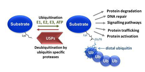
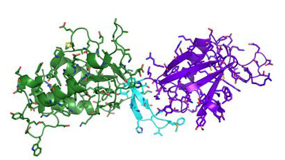
Figures:
Top: Schematic representation of ubiquitination and deubiquitination that regulates virtually all cellular events.
Bottom: Crystal structure of the ubiquitin specific protease 15 N-terminal interaction domains (PDB code 3T9L; Harper et al., 2011)
Project Aims:
In this project we will use structural biology techniques in combination with ligand binding assays to discover novel binding sites in these proteases that could be targeted by drug molecules. Furthermore crystal structures in complex with inhibitors will shed light onto how these could be modified for improved specificity. These structural insights into binding partner and inhibitor interactions will reveal fundamental principles of ubiquitin deconjugation, the molecular basis of signalling pathways implicated in disease processes and will be vital for the development of novel therapeutic agents targeting these enzymes. The project will provide training in a wide range of molecular biology, biochemical and structural biology techniques (including cloning, protein expression, enzymology and X-ray crystallography). Interested applicants are welcome to submit informal enquiries to ingrid.dreveny@nottingham.ac.uk.
Funding:
Sponsored or self-funded students of any nationality or students who wish to apply for their own funding are welcome to apply.
References
- Heideker, J., and Wertz, I.E. (2015). DUBs, the regulation of cell identity and disease. Biochem J 465, 1-26.
- Harper, S., Besong, T.M., Emsley, J., Scott, D.J., and Dreveny, I. (2011). Structure of the USP15 N-terminal domains: a beta-hairpin mediates close association between the DUSP and UBL domains. Biochemistry 50, 7995-8004.
- Harper, S., Gratton, H.E., Cornaciu, I., Oberer, M., Scott, D.J., Emsley, J., and Dreveny, I. (2014). Structure and catalytic regulatory function of ubiquitin specific protease 11 N-terminal and ubiquitin-like domains. Biochemistry 53, 2966-2978.
Additive biofabrication for regenerative medicine
Supervisor: Felicity Rose
Additive biofabrication is the use of three-dimensional (3-D) printing technologies to create structures that can direct the behaviour of biological systems. These structures are usually complex, and it is usually only possible to manufacture them with techniques based on additive 3-D methods. There is great interest therefore in applying these technologies to the field of regenerative medicine which aims to generate new treatments to enhance the tissue regeneration process following damage or disease. We also work with industry to provide in vitro human tissues that can be used to study disease or for toxicology/drug screening applications. The University of Nottingham is one of the world-leaders in additive manufacturing with state-of-the-art equipment and dedicated biofabrication laboratories (located in the Nottingham Biodiscovery Institute) to advance studies in this area. We can offer a number of projects in the area of additive biofabrication for regenerative medicine and welcome enquiries and PhD applications to work with us in these areas. We can only accept application from those that have their own funding or who are eligible to apply for scholarships.
Bioprinting of human tissue replacements for regenerative medicine and drug screening
Supervisor: Jing Yang
The field of bioprinting is undergoing an exciting period of development with the ultimate goal of producing tissues and organs suitable for transplantation. Several 3D printed cell-free medical devices have been deployed in the clinic. Making living tissues with long-term viability and functionalities is the next challenge to be addressed so that 3D bioprinting can benefit more patients. Several tissues including skin, bone, vascular grafts and cartilage have been made using 3D bioprinting, and some of them have been tested in animal models with promising results. This project will pursue the bioprinting of human tissues using printing techniques that are being developed in my laboratory. My group is currently working on a couple of human tissues including hepatic tissues and cartilaginous tissues which have potential for drug screening and transplantation respectively. Training will be given in all aspects of the project including 3D bioprinting, cell culture, immunostaining, microscopy (fluorescence, confocal, SEM), biochemical assays.
Exploring the cellular bioelectronic interface: Sugar mediated communication
Supervisor: Frankie Rawson
Following our recent discovery [1] that electron transfer occurs from eukaryotic cell surfaces of yeast and macrophage and is mediated via cell-surface saccharides it is our belief that this is ubiquitous to all eukaryotes. It is known that intracellular redox alterations play a role in cellular homeostasis. However, little is known regarding the function that redox modulations and bioelectronics interactions of cell-surfaces, and in particular, cell-surface sugars play in controlling cell function. The development and innovation of technology capable of forming a biocompatible interface between cells and materials in their environment is of great significance for an array of applications, from utilisation as a research tool to inform biological investigation through to cell culture to tissue engineering. The research proposed herein will elucidate the nature of the electronic surface interaction and how this modulates cell behaviour. This will allow us to delineate the biological role that cell-surface saccharide mediated redox changes play in modulating cell function, will seek to establish the ubiquity of this phenomenon and understand its mechanics. Thereby, offering a paradigm shift in the known role that cell-surface saccharides play in controlling cell homoeostasis.
[1]A. Stephenson-Brown, S. Yong, M. H. Mansor, Z. Hussein, N.-C. Yip, P. M. Mendes, J. S. Fossey, F. J. Rawson, Chem Comm, 2015.
Investigations into bioelectrochemical controlled fabrication of immune stimulating cell-surface bioflags
Supervisor: Frankie Rawson
We aim to develop a new synthetic bioelectrochemical-biology approach to label cancer cells with polymer “bioflags” enabling host immune systems to attack and kill diseased tissue more efficiently. There is a rapid need to develop non-invasive methods of labelling diseased tissues for a host of applications from treatment through to diagnostics. Saccharides are found on the surface of cells in the form of glycoproteins and glycolipids, in which sugars are sequentially attached to proteins or lipids (fats) to form biopolymers. These biopolymers play crucial roles in modulating cell function by acting as molecular flags. Therefore our approach will be to label cells with biopolymer mimics which, will be synthesized in real time on the surface of diseased cells
The function of polyadenylated long non-coding RNAs
Supervisors: Cornelia de Moor and Keith Spriggs
Long non-coding RNAs (lincRNAs) are a recently discovered diverse group of RNAs whose functions are largely unclear. Work in the De Moor group on mRNA polyadenylation has uncovered a group of polyadenylated RNAs that appear co-regulated on a number of conditions, such as growth factor stimulation, inflammatory stimulation and the inhibition of polyadenylation.
In this pioneering study you will use the latest molecular biology techniques (including quantitative PCR, immunoprecipitation, knockdown, thiouridine labelling, TAIL-seq, RNAseq) and advanced bioinformatics analysis to form hypotheses on the function of the polyadenylated lncRNAs and test them in tissue culture models of inflammatory diseases and cancer. This project is likely to lead to important fundamental discoveries on the function of a novel class of lncRNAs and ultimately to better treatment of cancer and/or inflammatory diseases.
For more information contact Dr Cornelia de Moor (cornelia.de_moor@nottingham.ac.uk)
The function of secondary metabolites from insect infecting fungi
Supervisiors: Cornelia de Moor and Dong-Hyun Kim
Cordycepin is isolated from Cordyceps militaris, a caterpillar fungus that is well known in traditional medicine. Recent work in the De Moor laboratory and elsewhere has shown that cordycepin has anti-inflammatory properties both in tissue culture and in vivo. It is likely that the fungus makes this compound to overcome the immune system of the caterpillars it infects. Indeed, injection of cordycepin reduces the induction of immune response genes by fungal spores in caterpillars. In this project, you will extract compounds from insect infecting fungi and examine their effect on the caterpillar immune system and the infection of caterpillars by fungi. You will use RT-qPCR, microscopy, RNA-seq and caterpillar observation to identify immune modulating compounds and investigate if their effects are also observed in mammalian systems. These studies will contribute to the development of novel biological pest controls and the discovery of novel anti-inflammatory drugs.
For more information contact Dr Cornelia de Moor (cornelia.de_moor@nottingham.ac.uk)
Finding the target of cordycepin in the PI3K/mTOR/AKT signal transduction pathway
Supervisor: Cornelia de Moor
The PI3 kinase pathway activates the mTOR and AKT kinases and is a well-known target for cancer therapy. The polyadenylation inhibitor cordycepin represses the PI3 kinase pathway by a so far unknown mechanism and has shown moderate activity against tumours in animal models. Recent work in the de Moor laboratory demonstrates that knocking down polyadenylation factors has a similar effect, indicating that PI3K/mTOR/AKT signalling requires polyadenylation. In this project you will use the latest techniques, such as polyadenylation assays, thermal proteome profiling and orthogonal organic phase separation (OOPS) to identify potential RNA and protein targets of cordycepin and subsequently use CRISPR/Cas9 knockout to confirm your undentification. Identification of the target of cordycepin will allow the development of more potent drugs working on the same principle.
For more information contact Dr Cornelia de Moor (cornelia.de_moor@nottingham.ac.uk)
Developing novel polyadenylation inhibitors as lead compounds for the treatment of cancer and pain
Supervisors: Cornelia de Moor and Michael Stocks
Polyadenylation is the last step of making a messenger RNA. We have shown that, contrary to the idea that all mRNAs are equally dependent on polyadenylation, growth factor induced and inflammatory mRNAs are much more sensitive to treatment with the polyadenylation inhibitor cordycepin. Indeed we have shown that cordycepin reduces breast cancer growth and relieves inflammation and osteoarthritis pain in animal models. However, cordycepin is relatively unstable and more potent inhibitors are required. In this project, you will develop two types of high throughput screens for novel polyadenylation inhibitors. One screen will be based on enzyme activity in a cell free sytem (biochemical assay), while the second one will be in tissue culture cells and use high content screening (microscopy). Initially you will test a small set of compounds to define the chemical space and then use modelling to select a library of other candidates. This project will discover novel lead compounds for the treatment of osteoarthritis pain and breast cancer.
For more information contact Dr Cornelia de Moor (cornelia.de_moor@nottingham.ac.uk)
Drug development and pre-clinical evaluation for Alzheimer's disease
Principle supervisor: Dr Zheying Zhu
Worldwide, approximately 50 million people are living with dementia, and this number is expected to reach 131.5 million by 2040. Alzheimer's disease is the leading cause of dementia and is thought to contribute to 60-70% of all cases. Its causes are thought as by multifunctional factors and the pathological mechanism is complex. To date, pharmacological therapies are very limited and not disease modifying treatments, therefore it has hugely unmet clinical needs.
There are a few relevant projects available in my group, including:
- The use of PROTACs technique to discover and develop novel drug candidates for Alzheimer's disease.
- Development of novel vaccines for Alzheimer's disease.
- The use of Drosophila melanogaster models to identify novel therapeutic agents for Alzheimer's disease.
- Gene based therapies for Alzheimer's disease and development of their delivery systems.
Sponsored and self-funded students are encouraged to contact Dr Zheying Zhu (zheying.zhu@nottingham.ac.uk) for further information.
Molecular and in-silico interrogation of beta adrenoceptor antagonists with a proposed novel binding mode
Supervisors: Shailesh N Mistry and Charles Laughton.
Background:
The beta adrenoceptor is a well-established drug target (“beta blockers”), but current molecules have poor selectivity between receptor subtypes. This limits the utility of these important drugs to patients with common co-presenting conditions – e.g. COPD. Through a recently completed PhD project we have identified, though advanced molecular modelling methods, a possible novel mode of binding of experimental subtype-selective ligands to beta adrenoceptors. This mode of binding may explain SAR data that otherwise appears contradictory.
The aim of this multidisciplinary project would be to design, synthesise and pharmacologically characterise a library of ligands to test this hypothesis more rigorously. In parallel, molecular modelling studies will be performed that eventually will be correlated with pharmacological data.
Main objectives:
- Design and synthesise a library of beta adrenoceptor ligands.
- Characterise the synthesised ligands pharmacologically, using a range of cell-based assays.
- Use advanced molecular modelling methods, including flexible docking and molecular dynamics simulations, to predict the mode of binding of new beta adrenoceptor ligands to the proteins.
- Make calculations of the binding affinities, and identify structure-activity relationships.
Major methods:
- Synthetic organic chemistry and structural elucidation.
- Cell culture and pharmacological evaluation of ligand affinity and subtype selectivity.
- Docking calculations using industry-standard molecular modelling software.
- Molecular dynamics simulations of protein-ligand complexes
- Post-processing of MD data to predict ligand binding affinities
Funding notes:
Applications are invited from self-funded students.
For informal inquiries please contact Dr Shailesh N Mistry (shailesh.mistry@nottingham.ac.uk) or Dr Charles Laughton (charles.laughton@nottingham.ac.uk).
Overcoming treatment resistance in pancreatic cancer
Supervisor: Dr Lodewijk Dekker
In spite of recent improvements, pancreatic cancer remains one of the most difficult to treat cancers. Current therapies include the use of the antimetabolite Gemcitabine. Development of resistance to this agent by the tumour cells is one of the factors associated with poor outcomes after treatment. Our laboratory has developed cell systems to model Gemcitabine resistance in pancreatic cancer cells. In this project we aim to understand how the cells can become resistant to Gemcitabine. Knowing this will help identifying novel ways to address the resistance and achieve better therapy outcomes.Preliminary data suggest that certain kinases that are implicated in regulation of the cell cycle may be activated in gemcitabine resistant cells. We will investigate this further using biochemical, pharmacological and genetic techniques. We will also employ a range of cell biological techniques that are widely used in cancer research. Overall, the project will provide thorough grounding in basic cancer research.
Investigating antitumour activities and mechanisms of action of novel molecules isolated from Malaysian Rainforest flora.
Supervisors: Tracey D Bradshaw, Kuan Hon Lim, John Moses.
In cross-campus collaboration with colleagues from University of Nottingham Malaysia Campus (UNMC) this project seeks to identify novel natural products which possess antitumour activity and elucidate molecular mechanisms of action. The multidisciplinary research encompasses tissue culture, pharmacology, microscopy, synthetic chemistry, analytical and `omics` techniques.
In recent projects, it has been shown that Jerantinine alkaloids isolated from Tabernaemontana corymbosa evoke antitumour activity 1,2 targeting tubulin, polo-like kinase 1 and spliceosome proteins. The crystal structure of Jerantinine B bound to the colchicine binding site of tubulin has been resolved. Using the Jerantinine skeleton, experiments will be undertaken to synthesise a small library of Jerantinine analogues to determine structure activity relationships (SAR).
Conopantinine, a structurally related but more complex molecule than Jerantinine, also isolated from Tabernaemontana corymbosa, has similarly shown potent antitumour activity, but via a distinct mechanism currently under investigation.
It is imperative that medicinal properties of flora indigenous to fragile habitats are investigated to help preserve such plants and identify potentially efficacious molecules. This is the goal of such research.
References
1. V. J. Raja, K. H. Lim, C. O. Leong, T. S. Kam and T. D. Bradshaw, Invest New Drug, 2014, 32, 838-850.
2. M. E. Qazzaz, V. J. Raja, K. H. Lim, T. S. Kam, J. B. Lee, P. Gershkovich and T. D. Bradshaw, Cancer Lett, 2016, 370, 185-197.
Applications are invited from self-funded students. For informal enquiries, please contact Dr. Tracey Bradshaw (tracey.bradshaw@nottingham.ac.uk)
Design, Synthesis and Evaluation of Novel Antitumour Pyrimethamine Derivatives
Supervisors: Shailesh N Mistry and Tracey D Bradshaw.
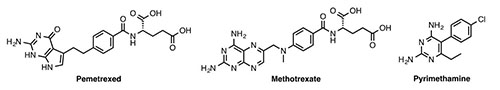
RAS is the most commonly mutated oncogene in cancer. Three RAS genes encode four proteins; KRAS is the most commonly mutated isoform found in pancreatic, colorectal (CRC) and non-small cell lung (NSCL) adenocarcinomas (among others). These mutations leave RAS in a permanently `on` state driving oncogenesis via various signal transduction cascades, enhancing proliferation and angiogenesis, disrupting metabolism and inhibiting apoptosis – seminal hallmarks of cancer (reviewed in Vasan et al, 2014).
KRAS mutations correlate with poor prognosis and resistance to chemotherapy and epidermal growth factor receptor (EGFR) inhibition in cancers such as NSCLC and CRC. Moran et al (2014) identified greater dependency on folate metabolism in KRAS mutant, compared with wild type (wt) NSCLC cell lines. Higher expression of genes in folate metabolism and purine synthesis-related pathways were detected in KRAS mutant cell lines. KRAS knockdown and overexpression studies demonstrated the ability of KRAS to regulate expression of genes within folate metabolism pathways; enhanced sensitivity to antifolates such as methotrexate and pemetrexed was found in KRAS mutant cells. KRAS gene expression was down-regulated by antifolates which may also contribute to enhanced activity of antifolates observed in KRAS mutant cells. In vivo, a pemetrexed-responsive, KRAS mutated tumour xenograft demonstrated an association between methylene-tetrahydrofolate dehydrogenase 2 expression and antifolate activity.
In the National Cancer Institute (NCI) 60-cell-line screen, growth inhibitory activity of novel derivatives of pyrimethamine with DHFR enzyme inhibitory activity, were also shown to exert activity via a non-folate mechanism of action. Exploitation of the computer pattern recognition algorithm (COMPARE) uncovered a positive correlation between activity and mutant KRAS in non-small-cell lung and colon cancer cell lines (Croughton et al, 2001). Modification of these lead structures may offer opportunities to generate novel molecules which target mutant RAS, an elusive protein target and malevolent driver of oncogenesis.
Project Aims
In this multidisciplinary project, the student will gain training in medicinal chemistry, organic synthesis, structural elucidation, and pharmacological and pharmaceutical skills pertinent to drug discovery. Specifically, the aims are to synthesise novel pyrimethamine derivatives, and to test the hypothesis that these molecules possess potent and selective antitumour activity, through a series of cell cytotoxicity and clonogenic assays. Activity of promising compounds will be examined in a panel of patient-derived NSCL, CRC and pancreatic tumour models characterised for expression of wt or mutant RAS.
References
Croughton et al, Anticancer Drug Des. 2001 16(2-3) 119-128. Moran et al, Mol. Cancer. Ther. 2014 13(6) 1611-1624. Vasan et al, Clin. Cancer. Res. 2014 20(15) 3921-3930.
Funding notes:
Applications are invited from self-funded students.
For informal inquiries please contact Dr Shailesh N Mistry (shailesh.mistry@nottingham.ac.uk) or Dr Tracey D Bradshaw (tracey.bradshaw@nottingham.ac.uk).
Structure and function of the platelet receptor Ib receptor. Opening doors to novel treatments for stroke
Supervisor: Jonas Emsley
Ischemic stroke is a devastating disease that represents the primary reason for sustained disability and the second leading cause of death worldwide. A better understanding of underlying molecular mechanisms of the disease are required to develop new treatment strategies. The interaction between platelet glycoprotein Iba (GpIbα) and von Willebrand factor (vWF) bound to damaged sub-endothelium represents the first step in platelet adhesion and is essential for normal hemostasis and vascular repair. The interaction is mediated between the N-terminal ligand-binding domain of GpIbα and the vWF-A1 domain and is markedly enhanced as hydrodynamic shear increases, due to conformational activation of vWF or GpIbα or both. In pathological situations, such as stroke or myocardial infarction, vascular damage and enhanced shear rates occurring in stenosised arteries can cause inappropriate activation, GpIbα-vWF binding contributing to thrombus formation. This project involves studying the molecular structure of the three proteins which form the GPIb complex (GPIba,GPIbb,GPIX) in complex with a key co-factor to coagulation enzyme and cell receptor function high molecular weight kininogen.
The Emsley group solved the first crystal structure of the GPIb N-terminal domain and an inhibitor complex (Blood.2009;114(23):4883-5). JE group subsequently described the structural organisation of the receptor and the molecular basis of the rare platelet disorder BSS (Blood.2011;118(19):5292-301). More recently through collaboration a peptide anti-GPIb approach was demonstrated in a mouse model of thrombosis (Blood.2014;124(25):3799-807). Experimental evidence suggests that GPIb is a critical pathogenic factor in innate immune inflammation in the brain. Ultimately the holy grail of my research is a greater overall understanding of GPIb assembly, regulation, and a more complete description of GPIb ligand and inhibitor interactions. This will provide a toolkit to open doors in establishing structure-function relationships and probe mechanism of brain disease (JournalofCerebralBloodFlow&Metabolism (2012)32,1831–1840)
http://www.nottingham.ac.uk/research/groups/structural-biology/index.aspx
Pharmacokinetics and pharmacodynamics of cordycepin, a potential lead drug in osteoarthritis
Supervisors: Cornelia De Moor and Pavel Gershkovich
Osteoarthritis is a common cause of pain and loss of flexibility in joints. It is characterised by a loss of cartilage, changes in the bone structure and synovitis in the affected joint. Treatment options are currently largely limited to exercise, non-steroid anti-inflammatory drugs, corticosteroids, opioid pain killers and surgery.
Cordycepin (3’ deoxyadenosine) is isolated from a fungus that is used as a health food and traditional medicine throughout the Far East. We have previously shown that cordycepin has anti-inflammatory properties through its effects on polyadenylation, the last step of mRNA synthesis. The specificity of the effect of polyadenylation inhibition on inflammatory mRNAs opens up the possibility that cordycepin could be a lead drug for the treatment of inflammatory disease.
The purpose of this project is to assess the biopharmaceutical properties of cordycepin, including its pharmacokinetics and biodistribution in order to evaluate the potential of cordycepin for the treatment of osteoarthritis.
Understanding where and how cordycepin is absorbed and how it distributes around the body is very important for the future development of better analogues with higher efficacy and low toxicity. This project will also help with the design of improved formulation and administration of unmodified cordycepin, e.g. it will indicate if intra-articular injection or a combination with a low dose of other compounds could improve the response.
Funding notes:
Applications are invited from self-funded students.
For informal inquiries please contact Dr Pavel Gershkovich (
pavel.gershkovich@nottingham.ac.uk) or Dr Cornelia De Moor (
Cornelia.De_moor@nottingham.ac.uk).
Targeting antiretroviral drugs to the gut-associated lymphoid tissues for improved treatment of HIV/AIDS
Supervisor: Pavel Gershkovich
Gut-associated lymphoid tissue (GALT) is an anatomical reservoir of HIV, setting a barrier for strategies aimed at HIV eradication. From a therapeutic perspective, limited data exist on the pharmacokinetics and pharmacodynamics of antiretrovirals within GALT. It has been proposed that incomplete immune reconstitution within GALT could be due to low levels of drugs within this compartment. Targeting the intestinal lymphatics is challenging because only a limited proportion of absorbed drug is distributed into the lymphatic system from systemic circulation. On the other hand, intestinal lymphatic transport following oral administration results in extremely high concentrations of the drug in the intestinal lymphatics. Therefore, the delivery of protease inhibitors to the intestinal lymphatics has potential to improve substantially the treatment of HIV/AIDS.
The aim of this PhD project is to improve the treatment outcomes of HIV/AIDS by targeting the antiretroviral drugs specifically to the GALT. This will be achieved by design and synthesis of prodrugs of protease inhibitors and other antiretroviral agents with high potential for intestinal lymphatic transport. Intestinal lymphatic transport is goverend by association of the molecules with chylomicrons in the enterocytes. An in silico model for association of drugs with chylomicrons will be used to evaluate the intestinal lymphatic transport potential of candidate molecules. Modifications of structures of protease inhibitors will be designed to yield prodruts that possess the appropriate physicochemical properties required for efficient intestinal lymphatic transport following oral administration. The purified produrugs will be assessed for the intestinal lymphatic potential by their degree of association with artificial chylomicrons-like emulsion (Intralipid®), and with natural chylomicrons separated from blood of human volunteers.
The pharmacokinetics and GALT distribution of the synthesised prodrugs and the corresponding active drugs will be assessed in vivo in a rat model. The antiviral activity of the drugs and prodrugs in the form that they appear in the GALT (associated with lipoproteins) will be assessed in vitro.
Finally, the in vivo efficacy of the propsed targeting approach will be assessed in a humanized mouse model of HIV infection.
Funding notes:
Applications are invited from self-funded students.
The project will be supervised by Dr Pavel Gershkovich and Dr Michael Stocks in the University of Nottingham, UK.
For informal inquiries please contact Dr Pavel Gershkovich (pavel.gershkovich@nottingham.ac.uk).
Targeting cannabinoids to the gut-associated lymphoid tissue (GALT) for improved treatment of autoimmune diseases
Supervisor: Pavel Gershkovich
Autoimmune diseases are conditions that are triggered by the immune system initiating an attack on the body’s own molecules. The causes of such attacks in most cases are unknown, but a number of studies suggest that they are associated with factors such as genetics, infections and environment. There are currently more than eighty different autoimmune diseases. Examples include debilitating conditions such as multiple sclerosis, systemic lupus erythematosus, arthritis and Grave’s disease.
There has been a long debate in the literature regarding the potential benefit of immunomodulatory effects of cannabinoids in the treatment of autoimmune diseases. These effects are based on the inhibition of immune cells (macrophages, T and B lymphocytes, natural killer cells) by cannabinoids via the cannabinoid receptor 2. Cannabinoids are highly lipophilic compounds. There are multiple reports that co-administration of lipophilic compounds with long-chain triglycerides (LCT) facilitates the intestinal lymphatic transport of these molecules. Lymphatic transport leads to extremely high local concentrations of lipophilic compounds in the lymph fluid and nodes. The delayed absorption of drugs when they are transported via the lymphatic system suggests that there could be several hours retention of cannabinoids in mesenteric lymph nodes following a single oral administration.
The rationale of this project is that oral administration of lipophilic cannabinoids with LCT facilitates the intestinal lymphatic transport and, thereby, creates high local concentrations of these molecules in the mesenteric lymph nodes and significant immunomodulation effect. We hypothesize that administration of lipophilic cannabinoids in conditions that facilitate lymphatic transport would result in a more efficient treatment of autoimmune diseases.
Over the last 3 years substantial in vitro and in vivo preliminary data have been generated in our laboratory. These data demonstrate the immunomodulatory activity of cannabinoids, as well as very significant intestinal lymphatic targeting with very high concentrations obtained in gut-associated lymphoid tissues (GALT).
Therefore, the aims of this PhD project will be focused on establishment of animal models of a number of autoimmune diseases, and demonstration of the efficacy of the GALT-targeting of cannabinoids in these animal models. The second half of the PhD will be focused on small-scale clinical studies that involve patients that suffer from the autoimmune disease that have been modelled in the in vivo studies.
Funding notes:
Applications are invited from self-funded students.
The project will be co-supervised by Dr Pavel Gershkovich, Professor Peter Fischer, Professor Cris Constantinescu and Professor Dave Barrett in the University of Nottingham, UK.
For informal inquiries please contact Dr Pavel Gershkovich (pavel.gershkovich@nottingham.ac.uk).
A novel alternative post-exposure prophylaxis local administration approach for prevention of HIV/AIDS
Supervisors: Pavel Gershkovich and Michael Stocks
HIV infections continue to be a heavy burden worldwide, with 36.7 million individuals living with HIV, and 2.1 million newly diagnosed individuals in 2015. Currently more than 80% of newly diagnosed infections are related to unprotected sexual intercourse with an HIV positive individual.
HIV is a virus that needs to integrate into the genome of TCD4+ lymphocytes in order to cause disease. In a sexual exposure scenario, HIV needs to travel through epithelium and find a dendritic cell which would transport it to a local lymph node, where TCD4+ lymphocytes can be readily found.
There are currently two main approaches in clinical use for HIV infection prevention based on antiretroviral drugs (in addition to main method of prevention using a physical barrier such as condoms):
- PrEP (Pre-exposure prophylaxis): This strategy is based on a low dose oral regimen of antiretrovirals. It is reserved for individuals with specific risk factors.
- One month high-dose PEP (post-exposure prophylaxis): This strategy is reserved for high risk contacts with an HIV+ source.
Current pharmacological approaches of HIV prevention leave low to moderate risk scenarios unaddressed. The aim of our work is to develop an alternative form of PEP which would be administered locally into rectum or vagina after potentially unsafe intercourse when risk is low to moderate.
Our hypothesis is that a nanoparticle-based drug delivery system would be distributed into the lymphatic system around the rectum or vagina (similar to the HIV virus itself), would saturate the system with antiretroviral agents, and therefore prevent the serum conversion.
Over the last several years we have assessed the need for such alternative prevention approach by means of socio-epidemiological survey involving more than 3000 responders. It was found that about 28% of sexually active population at least once in the last 3 years found themselves in such low to moderate risk scenarios, and therefore would be a target population for the alternative PEP approach.
Moreover, It is known that the absorption of rectally or vaginally administered nanoparticles is quantitatively low. Therefore, it is of paramount importance to achieve high drug loading in this novel nano-scale drug delivery system. We are now in a position in which we have a very high drug loading (above 50%) nanoparticle system with a first antiretroviral drug which we are about to test in vivo (in rodents) for local delivery to relevant lymph nodes after rectal administration.
This PhD project will involve optimization of the drug delivery system for local rectal and/or vaginal administration including loading with different antiretroviral agents and active molecular targeting to optimize uptake of the drug-loaded nanoparticles by relevant denritic cells and lymphocites. The research will also involve in vitro and in vivo experiments aimed to assess the distribution of the nanoparticles and release of the active drug in the relevant lymph nodes and relevant cells. This will also involve advanced imaging technologies to monitor the uptake fo the antiretroviral drugs by relevant cells. Finally, in the framework of this PhD efficacy studies of the novel PEP aproach will be performed in non-human primates with our collaborates in the USA.
Funding notes:
Applications are invited from self-funded students.
For informal enquiries please contact Dr Pavel Gershkovich (pavel.gershkovich@nottingham.ac.uk).
Targeting anticancer drugs in the intestinal lymphatic system for improved treatment of cancer
Supervisors: Pavel Gershkovich and Michael Stocks
The lymphatic system plays an important role in the pathophysiology of cancer as it can be both a site for the primary cancer and the major route of metastasis. Involvement of the intestinal lymphatic system is a predominant finding in 30-50% of the patients with non-Hodgkin’s lymphomas. Intestinal lymphatic system also provides the primary transportation route for the spreading of tumour cells from the gastrointestinal tract and other cancers from the pelvic region, therefore it is a critical pathophysiological compartment in cancer metastasis. It is challenging for systemic chemotherapy to specifically target the tumour in the lymphatics as only limited amount of drug can be distributed from the systemic circulation into the lymphatic system.
Discovery and development of antineoplastic agents with potential for intestinal lymphatic absorption following oral administration could significantly contribute towards improvement of survival rates of these patients. The distribution of drugs to the lymphatic system after oral administration is determined mainly by the association of drugs with large lipoproteins (chylomicrons) in the enterocytes. Therefore, by designing derivatives of anticancer agents that possess the necessary physicochemical properties for enhanced association with chylomicrons, we will be able to develop anticancer agents that target the lymphatic system. With chemical modification of existing or new anticancer agents, and lipid-based formulations that facilitate lymphatic transport, the concentration of such agents in the lymph can be achieved at levels of up to 500-fold higher than in plasma. Over the last several years substantial in vitro and in vivo preliminary data have been generated in our laboratory. These data demonstrate the efficient lymphatic targeting of anticancer compounds bexarotene and retinoic acid using prodrug approach.
Therefore, the aims of this PhD project will be focused on design and synthesis of prodrugs of additional (more novel and active) anticancer compounds, assessment of the intestinal lymphatic transport of these compounds, and assessment of the efficacy of the lymphatic targeting approach in cells and in animal models.
Funding notes:
Applications are invited from self-funded students.
For informal enquiries, please contact Dr Pavel Gershkovich (pavel.gershkovich@nottingham.ac.uk)
New treatments for tuberculosis
Supervisor: Cristina De Matteis
In collaboration with Professor Neil Thomas, School of Chemistry
Tuberculosis is one of the great unmet medical needs of present time, with estimates of 9 million people developing the disease and 1.5 million deaths annually across the world, and the rapid emergence of resistance to established treatments. This project aims to identify new drugs that are able to treat tuberculosis, including the resistant forms of the organism. It builds on our previous work in this area, where we have identified using virtual screening of compound libraries, a number of novel inhibitors of a known target enzyme in Mycobacterium tuberculosis, which show promising inhibitory activity in both enzyme kinetics assays and inhibition of Mycobacterium growth. Moreover, this target circumnavigates a known mechanism by which resistance is conferred, and hence provides the possibility of also being applicable to resistant forms of the organism. This project aims to identify new antibiotics for the treatment of tuberculosis, including resistant forms.
This project is interdisciplinary in nature and will include techniques in molecular modelling, 3D structural studies (e.g. crystallography), biological assays, and synthetic medicinal chemistry.
Advanced metabolite profiling methods: Application to human health and disease
Supervisor: Dr Dong-Hyun Kim
Since metabolites are end products of gene expression or metabolic activities of cells, monitoring metabolic changes reflects more accurately the responses of the cell or organism to gene expression and its environment. Therefore, the investigation of metabolites is most suitable for analysing the intracellular effects of various perturbations. This interesting and challenging project will involve the development of new metabolite profiling methodologies using state-of-the-art Ultra-High Performance Liquid Chromatography (UHPLC) linked to Orbitrap and ion trap mass spectrometry (MS), and/or ambient MS. The student (bioanlytical background preferred) will apply the novel methodology to selected cells, tissues or biofluids to identify metabolic pathways, which are targeted by various perturbations. In addition to MS analysis, the student will carry out subsequent statistical analyses using specialised software tools and identification of a complete profile of metabolites in the biological samples using authentic standards and databases. This novel methodology will then be applied to study specific diseases in order to monitor disease onset and progress, and to identify diagnostic biomarkers. These biomarkers can be used for new targets for chemotherapy and the development of new drugs.
Promoting wound healing and re-epithelisation through surface engineering
Supervisor: Morgan Alexander
Co-supervisor: Amir Ghaemmaghami (Life Sciences)
Epithelia cells play a key role in forming physical barriers that protect body against various physical, chemical, and microbial insults. Epithelial tissue’s diverse structural and functional properties depend on its location. For example, in organs with direct contact with the external environment such as the cornea, respiratory tract, skin, and digestive system, specialized epithelial cells maintain the equilibrium between the body and the surroundings. Loss of epithelial layer integrity and function is the cause and consequence of many pathological conditions including cystic fibrosis, inflammatory bowel disease, burns and chronic wounds. Epithelium tissues have a high renewal capacity, due to the presence of resident stem cells. However, in the case of deep injuries such as extensive burns and subsequent persistent infection due to loss of skin barrier function, a limit is reached where the epithelium self-healing cannot occur. Other conditions such as inflammatory diseases could also compromise the function and renewal capacity of the epithelial tissues.
There is a substantial interest in better understanding of different chemical and physical (topographical) cues that can promote the growth and differentiation of epithelial cells. Identifying such conditions will lead to better understanding of the biological events leading to epithelial growth and crucially could pave the way of more efficient treatments for conditions like extensive burns and chronic and non-healing wounds.
In this project we aim to use a high throughput screening approach to discover surface chemistries and topographies that support growth and differentiation of human skin and lung epithelial cells. Once identified we will investigate the signalling and molecular pathways that are activated (or inhibited) in epithelia cells in response to identified physical and chemical cues. This is a multidisciplinary project that provides training in cell biology, human skin and lung models, material microarray and high throughput screening technologies and a broad range of analytical techniques such as TOF-SIMs, imaging and flowcytometery. This project is suitable for candidates with an undergraduate degree (First or 2i) in biological sciences, chemistry or related disciplines. Postgraduate training (MRes or MSc) in a related area would be advantageous but not essential. The project will be supervised Dr Amir Ghaemmaghami (Faculty of Medicine and Life Sciences) and Prof Morgan Alexander (School of Pharmacy). Informal enquiries can be made to Dr Amir Ghaemmaghami (amg@nottingham.ac.uk) or Professor Morgan Alexander (morgan.alexander@nottingham.ac.uk). Applicants should send a detailed CV and a covering letter to Dr Amir Ghaemmaghami (amg@nottingham.ac.uk)
Further Reading:
- The development of a 3D immunocompetent model of human skin. Chau DY, Johnson C, MacNeil S, Haycock JW, Ghaemmaghami AM. Biofabrication. 2013 Sep;5(3):035011. doi: 10.1088/1758-5082/5/3/035011
- Engineering functional epithelium for regenerative medicine and in vitro organ models: a review. Vrana NE, Lavalle P, Dokmeci MR, Dehghani F, Ghaemmaghami AM, Khademhosseini A. Tissue Eng Part B Rev. 2013 Dec;19(6):529-43.
- Combinatorial discovery of polymers resistant to bacterial attachment. Hook AL, Chang CY, Yang J, Luckett J, Cockayne A, Atkinson S, Mei Y, Bayston R, Irvine DJ, Langer R, Anderson DG, Williams P, Davies MC, Alexander MR. Nat Biotechnol. 2012 Sep;30(9):868-75.
Structure and function of the platelet receptor Ib receptor. Novel treatments for stroke
Supervisor: Prof Jonas Emsley
Ischemic stroke is a devastating disease that represents the primary reason for sustained disability and the second leading cause of death worldwide. A better understanding of underlying molecular mechanisms of the disease are required to develop new treatment strategies. The interaction between platelet glycoprotein Iba (GpIbα) and von Willebrand factor (vWF) bound to damaged sub-endothelium represents the first step in platelet adhesion and is essential for normal hemostasis and vascular repair. The interaction is mediated between the N-terminal ligand-binding domain of GpIbα and the vWF-A1 domain and is markedly enhanced as hydrodynamic shear increases, due to conformational activation of vWF or GpIbα or both. In pathological situations, such as stroke or myocardial infarction, vascular damage and enhanced shear rates occurring in stenosised arteries can cause inappropriate activation, GpIbα-vWF binding contributing to thrombus formation. This project involves studying the molecular structure of the three proteins which form the GPIb complex (GPIba,GPIbb,GPIX) in complex with a key co-factor to coagulation enzyme and cell receptor function high molecular weight kininogen. http://www.nottingham.ac.uk/research/groups/structural-biology/index.aspx
Supervisor: Prof Jonas Emsley
Bradykinin is a potent peptide that acts as a vasodilator and an inflammatory mediator and is implicated in a number of diseases including cancer, stroke and high blood pressure. Bradykinin is found to be at raised levels in a wide range of clinical conditions such as inflammation leading to asthma, cardiovascular diseases, and the pathogenesis of severe acute respiratory syndrome coronavirus 2 (SARS-CoV-2) viral infection and COVID-19. Similarly, urokinase plasminogen activator receptor (UPAR) has been shown to be elevated in multiple inflammatory conditions including in the airways of asthma patients and during viral infection. Due to the role of bradykinin as an engine of inflammation antagonists have been suggested as promising drug candidates for inflammatory states but few medicines are currently available. The Emsley group has previously published the first complex structure of the co-receptor gC1qR in complex with a key component of the bradykinin producing machinery and proposed the first model for assembly of the gC1qR-uPAR complex [PubMed: 32559765, 29619369]. Here we propose a novel approach to investigate the gC1qR-uPAR structure using a variety of methods and target the gC1qR-uPAR-receptor complex.
Evaluation of new biodegradable polymeric nanoparticles for drug delivery to the lung
Supervisor: Dr.Cynthia Bosquillon
Polymeric nanoparticles could overcome many current limitations of drug inhalation. Unfortunately, most conventional polymers are not compatible with the pulmonary route of delivery due to concerns around their potential long-term accumulation in the lung tissue. The project will explore the potential of new green enzyme-synthesised polymers for efficiently delivering small or large therapeutic molecules to the lungs. This will include assessing nanoparticle characteristics, encapsulation efficiency, stability, biodegradability, interactions with cellular and non-cellular biological barriers in the lungs, etc…
For informal enquiries, please contact Dr. Cynthia Bosquillon (cynthia.bosquillon@nottingham.ac.uk)
Advanced responsive polymers for drug delivery
Supervisor: Professor Cameron Alexander
Materials that can respond to stimuli are of interest as carriers that can protect drugs during transport in the body and release the drugs where and when they are most needed. We are investigating new ways by which these materials can respond to biological signals and are developing polymers that can release drugs, biopolymers and complex macromolecular therapeutics for applications in cancers, neurodegenerative diseases, bacterial infections and as vaccines.
To apply
Please email Professor Cameron Alexander.
Synthetic polymer-cell interactions
Supervisor: Professor Cameron Alexander
The interactions of polymers with both bacterial and mammalian cells are important in disease processes and in regenerative medicine. We are looking at how synthetic polymers bind to cells in suspension and at surfaces, and future projects in this area will consider changes in cell biology (gene expression, protein and polysaccharide production) as a consequence of polymer-cell recognition.
PhD projects in both these areas are highly multidisciplinary, involving synthetic organic, medicinal and polymer chemistries combined with biophysical studies of polymer-biopolymer and polymer-cell interactions. Through collaborations with other groups we also evaluate drug, biopolymer and gene release efficacy in cell culture, and assess cell behaviour, signalling and gene regulation in response to polymer interactions.
To apply
Please email Professor Cameron Alexander.
Raman microscopy for characterising solid dosage forms
Supervisor: Dr Jonathan Burley
Drug and excipient distribution in tablets underpins their therapeutic performance. New developments in Raman microscopy/spectrometry allow a full and detailed chemical mapping of a tablet to be performed in 7-10 minutes. This project will involve looking at real-world examples, including problematic formulations, with a view to improving the design of old and new medicines.
Extreme miniaturisation for discovering new formulations
Supervisor: Dr Jonathan Burley
All drugs need to be formulated (made into a medicine) before they can be taken by patients. Current formulation practice requires time- and resource-intensive testing of several formulations in order to select the final optimised medicine. This process can take months and require significant quantities of often drug product, which is often in short supply, expensive and potentially quite toxic (e.g. cancer treatments).
Efforts to date in miniaturisation have employed scaled-down versions of lab-scale systems and are able to run individual experiments using milligrams of sample, typically sequentially. However these sample quantities are still problematic in terms of cost, availability and safety (many drugs exhibit biological effects at milligram and sub-milligram doses) and the experiments are still very time-consuming. In industrial development only 3-10 formulations can typically be tested using the current state of the art approach which is very limiting in developing a good understanding of likely product performance.
The project will look instead at "extreme miniaturisation", in which a new approach based upon 2D inkjet printing is used to create a nano-gram scale micro-array of multiple formulations which can be tested in parallel. The very low sample quantities used in the "extreme miniaturisation" mean that safety concerns for the formulating scientists largely disappear as the sample quantities used are orders of magnitude below any likely therapeutic level. The array approach (plus the extremely small amounts of sample required per test formulation) means that hundreds of samples can examined very rapidly. This offers new game-changing opportunities for rapidly, efficiently and cheaply developing many new formulations and the proposed project will look into several of these exciting openings.
Analytical approached will include: optical microscopy (and polarised light microscopy); Raman chemical microscopy; dynamic light scattering; X-ray diffraction; fluorescence microscopy; etc. Full training will be provided in these areas and also in the important area of high-throughput analysis of large data sets using open-source software such as "R" and "python".
To apply
Email your CV and interest to Dr Jonathan Burley.
Novel enabling chemical methods to macrocyclic peptides and their evaluation as antimicrobials
Supervisor: Dr Weng Chan
Unique naturally occurring macrocyclic peptides are important chemotypes for the exploration of new therapeutic agents for the treatment of bacterial infection. Inspired by naturally occurring templates such as argyrin and teixobactin, this project will entail the development of novel macrocyclic peptides. Using modern synthetic organic chemistry, new aromatic and aliphatic amino acid analogues/surrogates will be synthesised via chiral auxillary agents and palladium-catalysed cross-coupling reactions. These unique amino acid building blocks will then be incorporated into a Macrocyclic peptide framework, which will be assembled by modern solid-phase peptide synthesis strategies. These novel analogues will be extensively evaluated for their antimicrobial activities, with an aim to discover narrow-spectrum/pathogen-specific antimicrobials.
Understanding the Protein Kinase C delta pathway in disease
Supervisor: Dr Lodewijk Dekker
The Protein Kinase C delta (PKC-δ) pathway has been associated with various disease states, including inflammation and stroke and malignant diseases such as breast cancer. To understand underlying mechanisms, it is important to know the exact molecular targets of this kinase, which are currently poorly characterised.
We aim to identify PKC-δ-mediated phosphorylation events. A dominant negative strategy complemented by chemical genetics approaches will be employed to manipulate PKC-δ enzyme activity in cell models. Phosphorylated proteins will then be isolated and separated using chromatography and gelelectrophoresis. Proteins will be identified by means of mass spectroscopy methods. The results will lead to better understanding of the PKC-δ pathway in disease and potentially identify novel targets for pharmacological intervention.
mRNA metabolism as a target for anti-inflammatory drugs
Supervisor: Dr Cornelia H. de Moor
Chronic inflammatory diseases include afflictions such as rheumatoid arthritis, inflammatory bowel disease and asthma. In addition, inflammation has also been shown to play a role in several types of cancer and was recently linked to aging and coronary heart diseas. Therapeutic choices for inflammatory disease are limited to the non-steroid anti-inflammatory drugs (NSAIDS), which inhibit prostaglandin synthesis by cyclooxygenases, and corticosteroids, which act as repressors of inflammatory gene transcription. Both types of drugs have reduced effectiveness with long term usage, do not work in certain patients and can have serious side effects. Anti-inflammatory drugs that work on different principles could therefore significantly benefit a large number of patients with a variety of ailments.
Inflammatory gene transcription is mediated by the translocation of the transcription factor NFκB to the nucleus, which induces the transcription of inflammatory genes, including cytokines, chemokines, cyclooxygenases and transcription factors. The expression profile of these genes has recently been demonstrated to depend heavily on the stability of their mRNAs. Many of the key regulatory mRNAs were shown to be expressed transiently because of their instability, rather than due to transcriptional down regulation. Therefore, agents that specifically target these efficiently transcribed, unstable mRNAs could have a high potential as anti-inflammatory drugs.
In the De Moor laboratory, we have recently identified a new class of anti-inflammatory drugs that act at the post-transcriptional level. In this project you will use medium and high through put assays to identify more potent compounds working by this novel mechanism and study their effectiveness and metabolism in cells and animals.
How chromatin regulators read the epigenetic language
Supervisor: Professor David Heery
We study the structure and function of epigenetic regulators of gene expression using biochemistry, cell biology, chromatin IP, chip-Seq and structural biology. Several projects are available to study the structure and function of the Histone Acetyltransferases MOZ/MYST3 and MORF/MYST4. We recently solved the crystal structure of the MYST3 Double PHD domain in complex with histone tails (Dreveny et al, Submitted). Remarkably we discovered for the first time that MYST3 can induce a-helical structure of the tail which enables it to read and catalyse histone acetylation and methylation signals. Projects are available to study this domain structure in MYST4 and related proteins.
MOZ TIF2 function in acute myeloid leukaemia
Supervisor: Professor David Heery
MOZ/MYST3 is a histone acetyltransferase that is important for the development of haematopoietic stem cells. However, with recurrent chromosomal translocations in acute myeloid leukaemia patients express a fusion protein (MOZ-TIF2) which is sufficient to cause AML in animal models (Kindle et al., MCB 2005; Collins et al., JBC 2006; Kindle et al Leukaemia, 2010). We have used chromatin Immunoprecipitation coupled with Next Generation Sequencing techniques to identified gene targets of MOZ in the K562 cell line.
The project would aim to use RNA seq to identify genes that are misregulated by MOZ TIF2. The project will include molecular and cell biology, siRNA techniques, lentiviral expression of MOZ-TIF2 and NGS techniques. Some background in any of these techniques would be desirable.
Nuclear receptor/cofactor interactions
Supervisor: Professor David Heery
We have shown that the helical signature sequence LXXLL mediates interactions of Nuclear receptors and their many cofactors and that these interactions are essential for gene regulation (Heery et al., Nature 1997). Recent work from the lab has discovered novel highly selective sequences (FSXXLXXL) in some regulators that allow them to bind a subset of Nuclear receptors known as TLX, PNR and the COUP-TFs (Chan et al., Nucleic Acids Res 2013, In Press).
Several projects are available to investigate the structure and function of Nuclear receptor / Coregulators using, yeast two hybrid, proteomics and structural biology. For example now discoveries on the interaction of the retinal specific PNR are of potential relevance in understanding diseases involving sight loss via retinal degeneration. Other cancer-related projects under this theme also available.
Investigation of the factors required for regulation of hepatitis C virus by microRNA-122.
Supervisor: Dr Catherine Jopling
Hepatitis C virus (HCV) is a positive sense RNA virus that establishes persistent infections in the liver that may eventually result in cirrhosis or hepatocellular carcinoma. MicroRNAs are short, non-coding RNA molecules that are major regulators of gene expression in a broad range of eukaryotes. A liver-specific microRNA, miR-122, binds directly to hepatitis C virus (HCV) RNA and positively regulates viral replication. The mechanism by which this regulation occurs is not yet clear.
This project will investigate the protein factors required for the regulation of HCV by miR-122 in order to improve understanding of this important regulatory process.
Self-assembly of prodrugs for intra-tumoral delivery
Supervisors: Dr Maria Marlow, Professor Barrie Kellam, Dr Tracey Bradshaw
Securing the supply of medicines and understanding factors which can affect their performance.
Higher dose administration of a toxic chemotherapeutic directly into a tumour site (intratumoral delivery) is an obvious and alternative treatment to systemic delivery. Localised intra-tumoral delivery will reduce systemic exposure and hence reduce the potential for side effects. There is also the potential to form a depot for sustained release.
The Project will involve taking an existing chemotherapeutic and creating a prodrug that will self-assemble to form a physical gel in-situ upon injection into a tumour. The sol to gel transition will either be achieved by modification of the pH or temperature and could take advantage of the acidosis within the tumour. Introduction of a wide range of physicochemically distinct groups to metabollically liable tether points will be evaluated via parallel synthesis to afford a diverse library for gelation assessment. Once the conditions for gelation have been identified, the physical properties and bonding modes within the gels will be characterized. Enzymatic conversion of the prodrug to the drug will also be evaluated in vitro.
Finally the project will use an appropriate preclinical cancer model (e.g subcutaneous xengoraft) to evaluate the localised and systemic delivery. Research training will be broad based in that it will combine synthetic, physical chemistry, formulation science and cancer biology. This studentship will also provide an insight into designing and developing a drug product/therapy for oncology.
3D printing of solid dosage forms
Supervisor: Professor Clive Roberts
We have shown in recent years the ability of 3D printing to produce many types of solid dosage forms difficult to manufacture using current methods. This includes dosage forms with multiple APIs and release mechanisms. We have a number of major research programmes developing new printable formulation materials for solid dosage forms and implants. We now wish to exploit these formulations and develop robust scale up manufacture methods using 3D printing. You will work in a new facility dedicated to 3D printing of medicines in collaboration with engineers and clinicians.
Predicting the mass manufacturability of formulations based upon nano and micro scale measurements
Supervisor: Professor Clive Roberts
There is considerable interest in developing new strategies to select appropriate lead molecules based not only on their therapeutic profile but also the ability to develop them into a viable medicine that can be manufactured. Very many compounds fail at this point. This PhD will address this key issue.
The quantitative spatial and chemical analysis of formulations at the micron scale is now routine, using approaches such as infra-red and Raman spectroscopy and imaging. In recent years some nanoscale analyses using the probe microscopes have also become commonplace. This includes, for example the measurement of morphology, surface energy, elastic modulus and adhesion phenomena from nanoscale regions and frequently from single particles. You will build on this background to develop strategies for lead compound selection based upon nanoscale analysis.
We have developed a significant range of approaches and publication firsts in the application of nanoscale analysis of pharmaceuticals. Much of this work has been done in close collaboration with industry. You will employ a range of analytical techniques but in particular scanning probe microscopes, such as atomic force microscopy and nanothermal analysis.
Prospective candidates should have a good (2:1 or above) degree in a relevant subject area (e.g. pharmacy, materials science, physics, chemistry) and meet the University requirements for English (an IELTS score of 6 with no less than 5.5at each skill or 79 in the IBT TOEFL (with no less than 17 in listening and writing, 18 in reading and 20 in speaking). Alternatively, taking an appropriate pre-sessional English Course at the University’s Centre for English Language Education and achieving PASS in Academic English Level 2.
Design and development of paradoxical nanostructures for smart materials
Supervisor: Professor Phil Williams
It is possible to conceive materials that show paradoxical behaviour when stressed; springs that shorten when pulled, cylinders that thicken when stretched, etc. In this project we will use modern computational simulation techniques to design and refine molecular architectures that show such behaviours. This work will involve the use of the University’s multi-thousand processor high performance computer to undertake molecular dynamics simulations, bioinformatic techniques of sequence, structure and function database searching, and theoretical and analytical approaches to create generally applicable rules for the design of novel materials with unique properties. Such materials may have application in the creation of new types of sensors, fail-safe systems and mechanical devices, to name but a few.
Phylomechanics: The evolution of the mechanical properties of proteins
Supervisor: Professor Phil Williams
Proteins have evolved with specific function, some of which is mechanical in nature: cell adhesion proteins, muscle proteins, mechanoreceptor proteins all have to withstand mechanical stress. The mechanical properties of proteins have, therefore, evolved. Many questions can be asked of this evolution; what are the driving forces? Do force-bearing proteins share common ancestry? Can the effect of mutation be predicted? In this project we will develop our use of computational and bioinformatic tools for the rapid analysis of protein mechanical properties for the exploration of evolutionary links in mechanical function; phylomechanics. The project will apply computational tools being developed in our laboratories across protein structural databases using the University’s multi-thousand processor high performance computing systems. The outcomes of the project will increase our understanding of evolution, protein folding and degenerative disease.
Development of small-molecule inhibitors of enzymes involved in post-transcriptional gene regulation
Supervisor: Dr Sebastiaan Winkler
Potent, selective and cell-permeable inhibitors of enzymes are powerful tools for the study of protein function. Because such chemical probes share many characteristics with therapeutic drugs, they can play important roles in the validation of potential therapeutic targets and early-stage drug discovery.
This project will focus on the development of screening assays, and the subsequent identification of drug-like inhibitors of enzymes involved in post-transcriptional gene regulation. An example of an enzyme involved in this pathway is Caf1/CNOT7, a Mg(II)-dependent ribonuclease enzyme, which is a potential therapeutic target for the treatment of osteoporosis. The project will involve the expression and purification of recombinant proteins, automated screening of compound libraries, and the biochemical and cellular characterisation of drug-like inhibitors.
Role of BTG1 and BTG2 in acute lymphocytic leukaemia and diffuse large B-cell lymphoma
Supervisor: Dr Sebastiaan Winkler
Acute lymphocytic leukaemias (ALL) and diffuse large B-cell lymphomas (DLBCL) often contain small deletions of or point mutations in the highly related BTG1 or BTG2 genes. However, it is not clear how mutations in BTG1 and BTG2 contribute to tumourigenesis.
This project aims to gain further understanding regarding the contribution of BTG1 and BTG2 to tumour development. This will be investigated by knockdown of BTG1 and BTG2 mRNA in B cell lines in combination with the expression of mutant BTG1 and BTG2. The effects on gene expression as well as cellular proliferation and differentiation will be investigated by a variety of modern techniques, including real-time quantitative PCR, flow cytometry and fluorescence microscopy.
Gene based therapies for Down Syndrome and their delivery systems
Supervisor: Dr Zheying Zhu
Down Syndrome is the most common genetic neurodegenerative disease occurring in newborn babies, however, nearly all of the patients will develop Alzheimer's disease pathology starting from adults. Currently there is no effective therapies for the treatement of this condition, due to knowledge gaps in pathological mechanism, target identification, drug development towards the condition. With its twin promise of targeting disease aetiology and long-term correction, gene-based therapies are particularly appealing for neurodegenerative diseases, in particular Down Syndrome, where conventional pharmacologic approaches have been largely disappointing.
For more details of projects, sponsored and self-funded students are encouraged to directly contact Dr Zheying Zhu (zheying.zhu@nottingham.ac.uk) for further information.