The Childhood Ataxia Telangiectasia Neuroimaging Assessment Project (CATNAP)
What is CATNAP?
The CATNAP study aims to find MRI biomarkers of Ataxia Telangiectasia (A-T) in children.
A 'biomarker' is something that can be measured (for example in the blood or urine, or on a MRI scan) to monitor a biological process, disease or treatment response.
We will perform state-of-the-art magnetic resonance imaging (MRI) brain scans of healthy children and children with A-T. By comparing their MRI scans, we hope to find biomarkers which will be used in future studies to monitor new treatments designed for children with A-T.
Find out more about MRI below.
Contact
If you would like to participate in the study, or would like more information, please contact us at CATNAP@nottingham.ac.uk
More about participating in this study
Whatever your age, you can read more about participating in the CATNAP study using the following documents:
Magnetic Resonance Imaging (in short MRI) is a medical imaging device that uses a strong magnetic field and radio waves to create images of the inside of the human body. MRI does not involve any harmful radiation (e.g. X-rays) and so can be safely performed in most people.
MRI scanners allow us to look inside the human body with great detail. In the CATNAP study we will compare the size and shape of a part of the brain called the cerebellum (which is important for coordinating movements) in children with and without A-T.
MRI scanners can also be used to measure a number of biological processes.
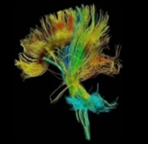 With diffusion imaging we can measure the microscopic movement of water in the brain. That information allows us to look at the brain structure and the physical connections between different parts of the brain. Together with functional imaging (see below) we will use this technique to see how the connections between different parts of the brain are affected in children with A-T.
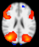 During different activities specific regions of the brain are used. With functional imaging we can show which brain areas are responsible for specific functions such as movement, speech or memory. The areas involved show up as orange segments and are using more oxygen than others during such tasks.
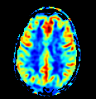 Perfusion imaging
 allows us to measure how much and how quickly blood flows to different areas of the brain. Any abnormalities related to anatomy, diffusion or brain function could help us to further understand the underlying neurodegeneration in children with A-T.
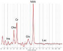 In MR spectroscopy the MRI scanner gives us a so-called MR spectrum instead of an image. A MR spectrum contains a number of peaks each of which belongs to a different chemical substance in the brain. With MR spectroscopy we hope to identify potential chemical imbalances in children with A-T. Such imbalances could be monitored in future treatment therapies to see if a treatment works.
[Back to Radiological Sciences research ]