Philips 7 Tesla Achieva
The laboratory is equipped with a 7 Tesla (7T) whole body multinuclear MR system (Philips Achieva) multi-transmit and receive capabilities.
This enables the lab to offer structural and functional imaging as well as spectroscopic measurement of energy stores and metabolite levels in the brain and body.
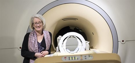
Professor Penny Gowland standing in front of the 7T MRI scanner
Capabilities
RF Coils
We have the following RF coils:
- 2 channel multi transmit Nova head coil
- 16 channel Nova head receive coil for use with above transmit coil
- 32 channel Nova head receive coil for use with above transmit coil
- TR Head Coil
- 8 channel transmit/ 32 channel receive body array coil from MRCoils
- A quadrature carbon coil with quadrature proton decouple coils (for 13C imaging and spectroscopy in muscle and brain)
- Proton coils (as used for imaging)
- A variety of other specialist coils, some developed locally
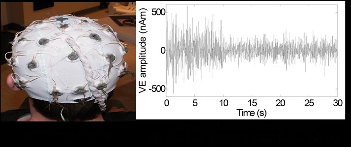
Electroencephalography (EEG)
32 or 64 channel MR compatible EEG system and caps (Brain Products GmbH). This allows functional MR and EEG signals (which measure neuronal oscillations directly) to be acquired simultaneously. Research in this area ensures advice can be given so that acquisition methods to minimise artefacts are optimal.
Electromyography (EMG)
EMG cap be recorded with unipolar or bipolar MR compatible amplifiers (Brain Products GmbH). This allows any muscle activity to be monitored which has a number of uses in functional imaging.
Physiological signals
Breathing, pulse rate and cardiac trace can be monitored using the Philips monitoring system.
Stimulus Presentation
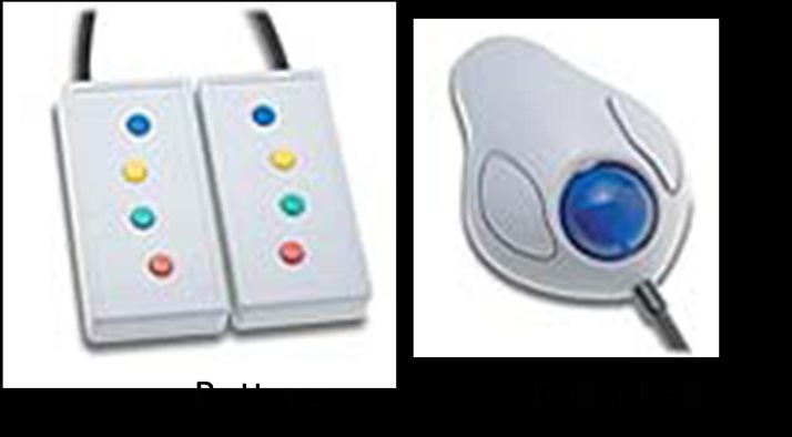
The lab is equipped to deliver most forms of stimulation including: visual (using goggles or projection to a screen), auditory, somatosensory (including median nerve and vibrotactile stimulation), taste and pain (temperature induced). A separate stimulus computer is used to present paradigms to the subject and this is equipped with MATLAB, PsychoPy and Presentation (Neurobehavioural systems) for paradigm generation. The lab also has equipment to monitor responses during a task using a fORP system. Currently available in the lab are buttons or a roller-ball but other inputs could be added.
The lab has MR compatible glass that can correct vision of +/-6 to assist people when performing tasks which require visual input.
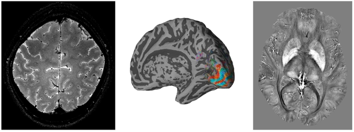
High resolution anatomical and functional brain images produced on the 7T scanner