EEG-fMRI: data quality Questionnaire Findings
In June-July 2019, we (Dr Karen Mullinger and Prof Richard Bowtell, SPMIC, University of Nottingham) under-took conducting a survey to better understand whether researchers felt they were facing EEG data quality limitations when using simultaneous EEG-fMRI as a research tool. We asked further questions to understand which of the EEG artefacts were deemed most problematic for their specific research interests and if hardware solutions were available what the practical limitations in terms of using these solutions would be.
We contacted 100 PIs and senior researchers worldwide who were identified as having used EEG-fMRI in their research (through their publication record). We achieved a 40% response rate. These responses are summarised below for each question separately. We interpret these results as showing:
- EEG data quality is still not good enough (73% of respondents have not been able to answer questions they wanted to)
- A broad range of frequency bands are of interest when using EEG-fMRI
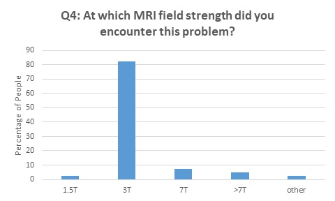
- Most (83%) people are using 3T MRI for their simultaneous EEG-fMRI experiments
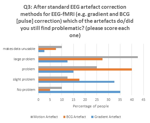
- The motion artefact is now the greatest problem for data quality, although people still have problems with the pulse and gradient artefacts
- 58% of people would be willing for set up to take 20 minutes longer or more (38% of people would take as long as needed) if hardware existed which could improve the data quality to the level required
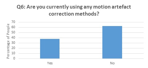
- Most people are not performing any type of EEG motion artefact correction and those who are, are using a wide variety of methods


Only those who answer yes to question 6 went on to answer question 7.

In question 7, MPT refers to the methods outlined by [1, 2], wire loops refers to methods outlined by [3-5], MR motion correction refers to methods where the MR data itself is used to detect motion although I believe this may not be used in EEG motion artefact correction and is hard to tell from responses. Reference layer refers to any method where a second conductive layer is used to measure the EEG artefacts such as [6-9].
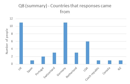
In total responses came from 30 different institutions, with different groups within some institutions answering separately.

References
1. LeVan, P., et al., Neuroimage, 2013. 75: 1-11.
2. Maziero, D., et al., Neuroimage, 2016. 138: 13-27.
3. Abbott, D.F., et al., Front Neurol, 2014. 5: 260.
4. Jorge, J., et al., Neuroimage, 2015. 120: 143-53.
5. Masterton, R.A., et al., Neuroimage, 2007. 37: 202-11.
6. Chowdhury, M.E.H., et al., Neuroimage, 2014. 84: 307-319.
7. Luo, Q.F., et al., Journal of Neuroscience Methods, 2014. 233: 137-149.
8. Steyrl, D., et al., Journal of Neural Engineering, 2017. 14.
9. Xia, H.J., et al., Frontiers in Neuroscience, 2014. 8.