Sense of Touch
The Skin and its Receptors
The skin is the largest organ in our body1, and is made up of two types: glabrous and hairy. These contain different types of mechanoreceptors, specialist mechanosensitive sensory organs, which receive and transmit a neural signal to our brain. Our group is interested in LTMRs (low-threshold mechanoreceptors), since these react to innocuous stimulation, such as non-painful touch.
Cutaneous (things related to/affecting the skin) sensory receptors are further subdivided into Aβ, AΔ, and C fibres. These fibres have different conduction velocities, cell sizes, axon diameter, and their degree of myelination. These are present different skin types, different regions of the skin and are sensitive to different stimuli, e.g. Aβ fibres have the fastest conduction velocity, can be present in both glabrous and hairy skin, and are sensitive to indentation, stretch, vibration and skin movement (in their subtypes).
The firing patterns are also different for mechanoreceptors, divided into slow-adapting (SAI/II) and rapidly-adapting (RAI/II). SA receptors exhibit constant firing to sustained indentation, whilst RA receptors fire only at the initial and terminal contacts of a mechanical stimulus.
More information on the skin and its receptors
Whilst SA eceptors are better suited to maintained stimulation, RA receptors are better suited to objects moving quickly across the skin.
Have a look at Table 1 for a comparison of cutaneous mechanoreceptors, and the four main types of LTMRS: Meissner corpuscles, Pacinian corpuscles, Ruffini endings and Merkel cells (which are all only present in glabrous skin)2.
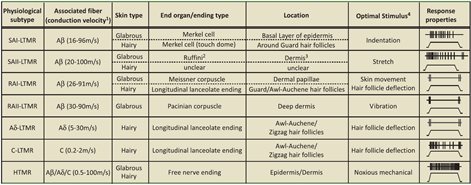 Table 1. A comparison of cutaneous mechanoreceptor types2
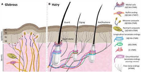 Fig2. The different receptors present in glabrous and hairy skin2

Fig1. The Hulk. He has a lot of skin.
Affective Touch
We have so far only described those sensory fibres which provide discriminative touch, but recently, a new set of fibres have been investiaged, which are thought to be related to affective or emotional touch3. These CT fibres are only present in hairy skin, and respond preferentially to soft brush stroking at velocities of 1-10 cm s-1, perceived as being pleasant by participants4. Projections to insular cortex (a site of emotional processing), and correlations with affective modulation/hedonic responses have led to several groups suggesting that these CT fibres are activated during 'affiliative interactions between conspecifics, constituting a peripheral pathway for gentle, pleasant tactile stimulation of a social nature.'
Pathways
The aforementioned afferent sensory neurones in the peripheral nervous system typically have their cell bodies in the dorsal root ganglion (in the spine); these pseudo-unipolar neurones will synapse with a secondary neurone travelling up the spinal cord, crossing and terminating in the thalamus (the brain's major road junction). The next synapse with a tertiary neurone ends up in the postcentral gyrus, the base of the somatosensory cortex5.
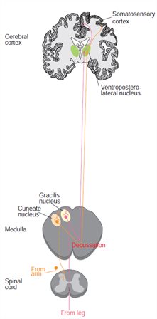
Fig3. The pathway from sensory neuron to S1
Electrophysiological findings
Studies performed in the late 70s and onwards have provided a wealth of information regarding the structure of S1, through invasive electrophysiological mapping, usually performed on monkeys. The cytoarchitectonic delineations of our brain were mapped by Brodmann, and areas 3a, 3b, 1 and 2 have been associated with S1 and S2.
Hierarchal descriptions, differing receptive field sizes6 and mirroring representations7 have littered the field of research, however, these interpretations have led to controversy; purely mapping studies are performed less (understandably) in animals, and it may be some time before non-invasive measures (such as fMRI) catch up to the quality of electrophysiological investigations and also catch up to the ability to answer those questions posed 40 years ago.
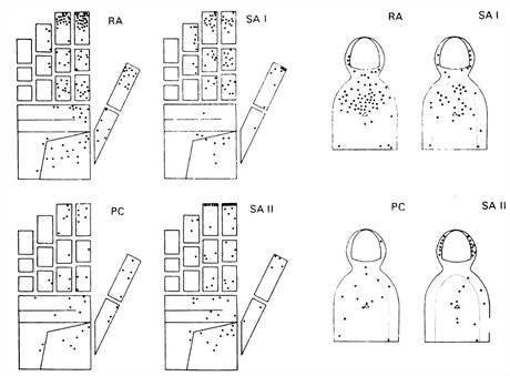
Fig4. Density and loci of various mechanoreceptors on the hand and fingertips, indicated by firing pattern subtype. Vallbo et al. (1979)
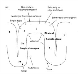
Fig5. The 'omega' shaped S1, seen in sagital view, indicating Brodmann areas7
References
- Fridman, I. Surface Area of Human Skin. (2001). at <http://hypertextbook.com/facts/2001/IgorFridman.shtml>
- Abraira, V. E. & Ginty, D. D. The Sensory Neurons of Touch. Neuron 79, 618–639 (2013).
- McGlone, F. et al. Discriminative touch and emotional touch. Can. J. Exp. Psychol. 61, 173–183 (2007).
- Löken, L. S., Wessberg, J., Morrison, I., McGlone, F. & Olausson, H. Coding of pleasant touch by unmyelinated afferents in humans. Nat. Neurosci. 12, 547–548 (2009).
- Squire, L. R. et al. Fundamental Neuroscience. (Elsevier, 2008).
- Kaas, J. H., Nelson, R. J., Sur, M., Lin, C. S. & Merzenich, M. M. Multiple representations of the body within the primary somatosensory cortex of primates. Science 204, 521–523 (1979).
- Iwamura, Y. Hierarchical somatosensory processing. Curr. Opin. Neurobiol. 8, 522–528 (1998).