Our Research
Figure 1: Panel showing single EPEC bacterium in wide field and Structured illumination and wide field and dSTORM.
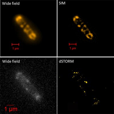
Figure 2: Epec bacteria labelled for cell wall and DNA.

Figure 3: Bovine Pulmonary Epithelial Cells labelled for actin (green), mitochondria (red) and nuclei (blue). Top panel confocal and SIM. Bottom panel cropped region from one of the cells with additional dSTORM image.

Figure 4: Melanocyte cell labelled for actin and myosin Va in wide field and SIM.

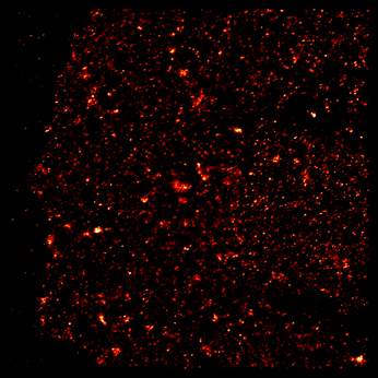
Figure 5: Adenosine receptor clustering in membrance of CHO cells with dSTORM.