Expertise and Equipment in the Regenerative Medicine and Cellular Therapies Division
The Division of Regenerative Medicine and Cellular Therapies is based within the Nottingham Biodiscovery Institute and the Boots Science Building. Our laboratories are well-equipped with cutting edge tools and technologies for scientific research in the manipulation and control of mammalian cells and biomaterials development.
3D Bioprinting
3D bioprinting is a burgeoning field that aims to produce functional human tissues and organs for tissue engineering and regenerative medicine. 3D bioprinting allows the spatial positioning of cells, biomolecules, and biomaterials within three-dimensional constructs. Our interests focus on developing novel printable biomaterials and printing approaches, and investigating the interactions between printed biomaterial scaffolds and cells/tissues in vitro and in vivo. We have various 3D printing technologies ranging from extrusion-based to light-based techniques. A range of biomaterials has been investigated including polymers, hydrogels, bioactive glasses and bioceramics.
Lead Investigator: Dr Jing Yang
Holographic Tweezers for Cell Manipulation
When a laser beam is shone into a liquid in which small objects are suspended, it is possible to pull those objects towards the beam. This phenomenon is known as optical trapping. By shining a laser beam on to a hologram, multiple laser beams are created which gives this phenomenon a third dimension. Once the object is trapped by the beam, it can be moved very precisely by simply controlling the laser via a joystick or touch screen linked to a computer system. These small objects can include mammalian cells and therefore this system provides a unique way of building very precise multi-cellular configurations or structures in vitro.
Lead Investigator: Dr Lee Buttery
Decellularised Extracellular Matrix Hydrogels
The extracellular matrix (ECM) of mammalian tissues has been used as a scaffold, following decellularisation, for the repair and reconstruction of a number of tissues. We have developed methodologies to produce decellularised scaffolds from bone. In addition, we have produced a soluble form which can be induced to form hydrogels with distinct structural, mechanical and biological properties. We are studying these hydrogels for their potential for clinical delivery and their potential to support tissue regeneration.
Lead Investigator: Dr Lisa White
Non-Viral Gene Delivery
Glycosaminoglycan-binding enhanced transduction (GET) is a peptide-based system engineered to enhance the activity of cell-penetrating peptides to achieve exceptional intracellular transduction. This technology uses peptides that interact with cell membrane heparan sulphates and promotes cell-penetrating peptide-mediated endocytosis without affecting cell proliferation and viability. This method of delivery is not dependent on extensive positive charge and can be tailored to deliver peptides, recombinant proteins, nucleic acids, nanoparticles, and antibodies. We have exploited this technology for gene correction, editing, infectious disease vaccines (COVID19 and Zika) as well as several regenerative medicine applications
Lead Investigator: Dr James Dixon
Design and Characterisation of Biomaterials and Biointerfaces
Biomaterial design supports healthcare related research for medical devices, drug delivery, disease models and regenerative medicine. We are exploiting chemical and mechanical functionalities of peptides, polymers and self-assembled materials on surfaces, at interfaces and in bulk hydrogels for controlled drug delivery and to direct cell behaviour and cell fate. We have expertise in N-carboxy anhydride polymerisations, peptide modification and tuning mechanical properties of gel films. Biomaterial and Biointerface characterisation is an integral part of our work. We routinely apply state-of-the art surface and interface analytical tools such as X-ray photoelectron spectroscopy (XPS), Time of Flight Secondary Ion Mass Spectrometry (ToF-SIMS) and atomic force microscopy (AFM) and we are developing ways to measure complex biological environments to allow us to develop an understanding of the cell-material interface where low concentrations of biomolecules may be present and surfaces may be hidden.
Lead Investigator: Dr Mischa Zelzer
Electrospinning
Electrospinning is a method of producing both nano- and micro-fibres from natural and synthetic polymers that replicate the morphology of the extracellular matrix. Our research laboratories house an IME technologies climate controlled electrospinning apparatus. Using electrospinning as a core technology, we have interests in the functionalisation of these scaffolds with chemistries to support and influence cell growth and differentiation. Using co-axial electrospinning, we are exploring the incorporation of bioactive molecules for controlled release. Application of electrospun scaffolds for mucosal tissue engineering, including gut, cornea, lung, and for skin wound healing is of particular interest. We also have interests in developing these materials for the 3D cell culture market.
Lead Investigator: Dr Felicity Rose
Controlled Release of Biopharmaceuticals for Tissue Regeneration
Biomolecules, such as growth factors, are important in the regulation of stem cell differentiation and tissue formation. Their delivery to the body requires the development of controlled release strategies such that physiologically relevant doses are delivered over a sufficient period of time to support tissue regeneration. In addition, for tissue engineering applications, there is a need to deliver more than one growth factor, for example to stimulate angiogenesis alongside the promotion of osteogenesis. We have invented numerous new delivery systems for the delivery of growth factors and cells and applied these systems in the regeneration of bone, cartilage, liver, nerve and corneal tissue.
Lead Investigator: Dr Lisa White, Professor Felicity Rose and Dr Jing Yang
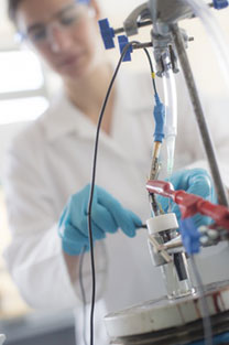
Bioelectronic sensors and actuators
We are entering an era where bioelectricity is a key dogma in biology. Malfunctions in these bioelectrical relays underpin disease manifestation. Consequently, to aid our understanding and modulating cell behaviour requires development of bioelectrical sensors and actuators that modulate cell function. We are developing new nanobioelectronic technologies to treat disease and manipulate cell metabolism. Allowing for both the sensing and actuating of novel cellular electrical relays with the aim of understanding and treating disease with electroceuticals and developing sustainable energy platforms. The groups work is multi-disciplinary and involves 3D printing of bioelectronics, electroanalytical chemical techniques including amperometry, potentiometry and impedance spectrometry, and cell molecular biology in understanding how bioelectricity underpins disease. We are based in the bioelectronics laboartor that has a suit of Autolabs and Versastat potentiostats with low current modules, wave generators and high voltage stimulators.
Lead Investigator: Dr Frankie Rawson
Image: Postgraduate student breaking down molecules using potentiostat equipment
c
Supramolecular biofabrication
As the need for more efficient regenerative solutions increases, it is essential to establish new fabrication approaches that can more accurately recreate these complex biological systems. We are developing innovative fabrication processes capable of assembling biomolecules hierarchically facilitating communication with biology at sub-cellular, cellular and multicellular levels. For example, by integrating additive manufacturing and self-assembly, we are growing structures by simultaneously printing from the top-down and self-assembling from the bottom-up. In another example, we use engineering principles to control organic-inorganic interactions to guide mineralization across size-scales and into materials with bioinspired properties.
Lead Investigator: Prof Alvaro Mata
Image: Peptide-protein co-assembly into vascular like capillaries