2.5 Low energy electron diffraction
References
Electron diffraction is a key technique used in elucidating the structure of surfaces. X-ray diffraction, as very commonly used for bulk structure determination may also be applied to surface studies but the experiments are much more difficult than the corresponding bulk investigations.  Can you give a reason why X-ray diffraction is difficult to apply to surfaces?
Surface electron diffraction techniques rely on the interference of electron waves elastically scattered from the surface of a crystal. The interference gives rise to Bragg diffraction spots whose positions are directly related to the periodicity of the surface.
There are two primary forms of electron diffraction used in surface analysis - low energy electron diffraction (LEED) and reflection high energy electron diffraction (RHEED). We will discuss LEED now and return to RHEED in Section 6.
A brief, though important, aside: I should point out at this stage that surface science is unfortunately
associated with a very large number of acronyms which are used for various techniques and processes. You’ve met three already : STM, LEED and RHEED and will meet many more before completing this module! A listing of acronyms and their meanings can be found here . This excellent site also gives a brief description of each technique
An interesting site which discusses the history of LEED (the first LEED experiment also provided the first demonstration of the wave nature of the electron!) is found here .
LEED apparatus
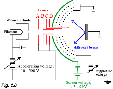
 A typical LEED apparatus is shown in Fig. 2.8 (adapted from Luth Fig VIII.1, p.228). Electrons are thermionically emitted by the heated filament and collimated by the Wehnelt cylinder (W) which has a small negative bias with respect to the filament. The energy with which the electrons are incident on the sample is determined by the acceleration voltage i.e. the voltage between the filament and apertures A and D in Fig. 2.8. Apertures B and C are used to focus the electron beam.
The incident electrons are backscattered from the sample surface and strike the fluorescent screen to create the diffraction pattern. The screen is at a high positive potential with respect to the sample surface so that the low energy, elastically scattered electrons are accelerated onto the screen.
There are also a number of grids before the screen. As is clear from Fig. 2.8, apertures A and D, two of the grids before the fluorescent screen and the sample are all grounded. Thus, a field free region between the sample and the screen exists - this is important to prevent electrostatic deflection of the electrons. Finally, a third grid is used to suppress the electrons that have been scattered inelastically from the surface. These electrons do not produce diffraction spots but contribute a uniform background illumination.
Some photographs of LEED patterns may be found via the link provided below. At the centre of each photograph you will see the LEED "gun" - the unit used to produce, collimate and focus the electrons. Around the gun in each photo the spots of the diffraction pattern are clearly visible. Click here to see the LEED patterns. In the next section we’ll discuss just how LEED patterns are related to the structure of the surface………..
Move on to Section 2.6: Reciprocal Space and Surfaces
Home
 A typical LEED apparatus is shown in Fig. 2.8 (adapted from Luth Fig VIII.1, p.228). Electrons are thermionically emitted by the heated filament and collimated by the Wehnelt cylinder (W) which has a small negative bias with respect to the filament. The energy with which the electrons are incident on the sample is determined by the acceleration voltage i.e. the voltage between the filament and apertures A and D in Fig. 2.8. Apertures B and C are used to focus the electron beam.
The incident electrons are backscattered from the sample surface and strike the fluorescent screen to create the diffraction pattern. The screen is at a high positive potential with respect to the sample surface so that the low energy, elastically scattered electrons are accelerated onto the screen.
 Can you give a reason why X-ray diffraction is difficult to apply to surfaces?
Surface electron diffraction techniques rely on the interference of electron waves elastically scattered from the surface of a crystal. The interference gives rise to Bragg diffraction spots whose positions are directly related to the periodicity of the surface.