What's at the nmRC?
In addition to acting as a learning and research hub for nano-, micro- and materials science at the University of Nottingham, the nmRC hosts a number of state-of-the-art research facilities.
The centre is a specialist resource for electron microscopy variants, with vast Transmission Electron Microscopy (TEM), Scanning Electron Microscopy (SEM), and electron microprobe infrastructure and dedicated technical support.
In addition the centre is also home to X-Ray Photoelectron Spectroscopy (XPS), Confocal Raman Microscopy, Secondary Ion Mass Spectroscopy (SIM) and sample preparation suites. Please follow the links below for more information or additional technical information can be found on the nmRC Commercial Services (nmCS) website, or is internally available to University staff and students via the Kit Catalogue.
We also have a selection of e-leaflets for most of our equipment which highlight key capabilities, instrumentation, and relevant case studies, which can be found at the bottom of the page.
Electron Microscopy
The nmRC is proudly home to a collection of world leading electron microscopy expertise, infrastructure and personnel. With three Transmission Electron Microscopes (TEMs), eight Scanning Electron Microscopes (SEMs) and an Electron Microprobe the centre offers a diverse range of electron microscopy specialisms to support research in a number of fields and disciplines.
Raman spectroscopy is a non-invasive optical spectroscopy technique used to determine the chemical identity and state of a sample by its unique vibrational modes. The inelastic scattering of light from the sample is measured, with shifts in the energy of the scattered photons being representative of the sample chemistry.
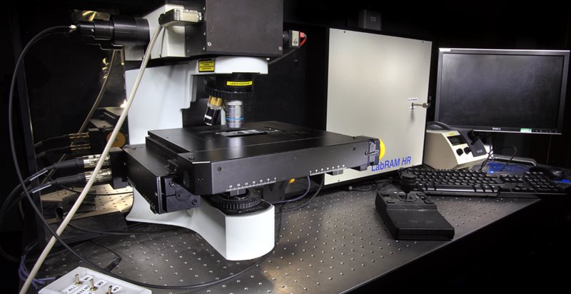
Secondary Ion Mass Spectrometry (SIMS) is a highly sensitive surface analytical method that can describe the chemical character of a substrate surface in 3D. Solid surfaces can be analysed by either a high mass resolution Orbitrap analyser or high imaging resolution time of flight (ToF) analysers.
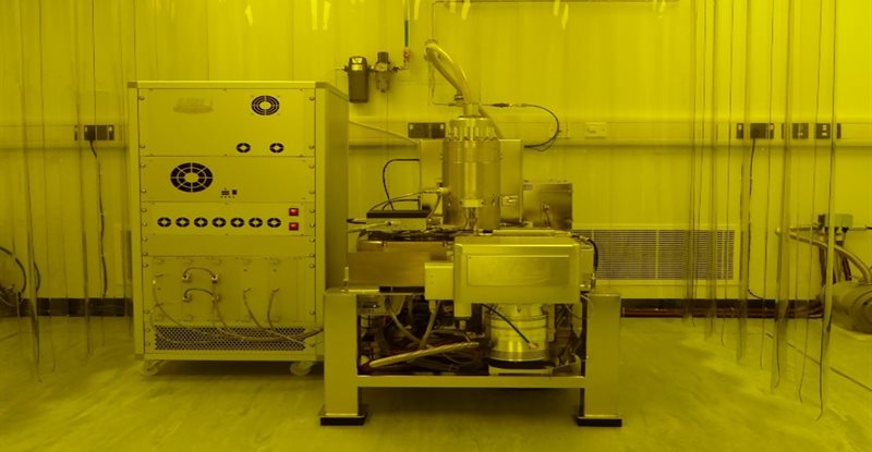
The Accurion nanofilm_ep4 imaging elipsometer allows for precise measurement of thickness and optical properties of thin films. Unlike conventional elipsometry, imaging elipsometry is able to produce maps of a sample showing how these properties vary across the surface.
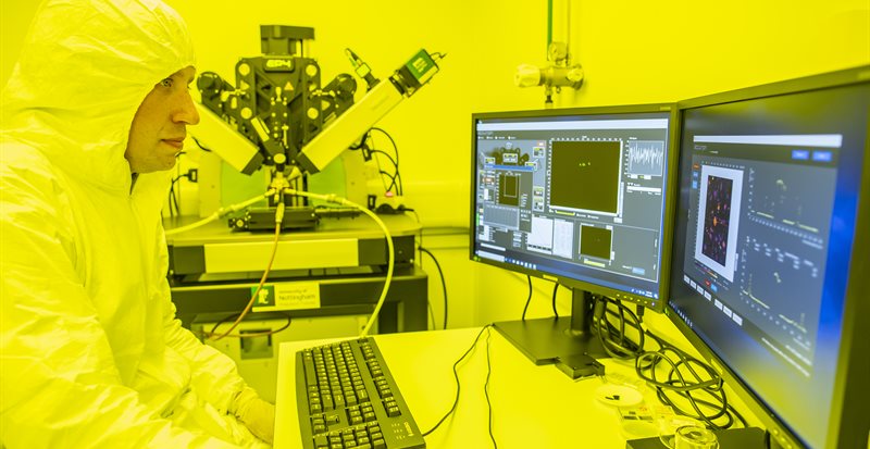
The Nanofabrication Nottingham (NaNo) cleanroom suite within the nmRC has been designed to perform high quality electron beam lithography (EBL) using a nanobeam nB5 EBL tool. EBL has been used in work in numerous disciplines from: physics to chemistry to biology and electrical engineering.
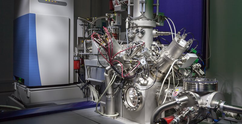
XPS is a surface sensitive chemical analysis technique that provides quantitative elemental characterisation of a sample. The nmRC is home to two multiuser XPS instruments equipped for versatile and multi-disciplinary research or commercial applications.
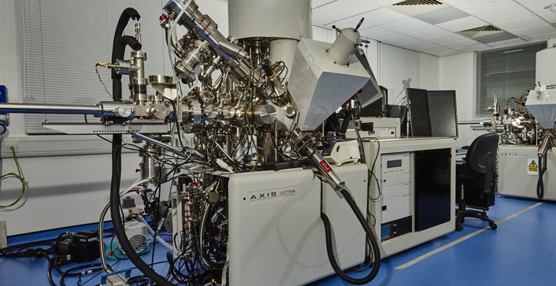
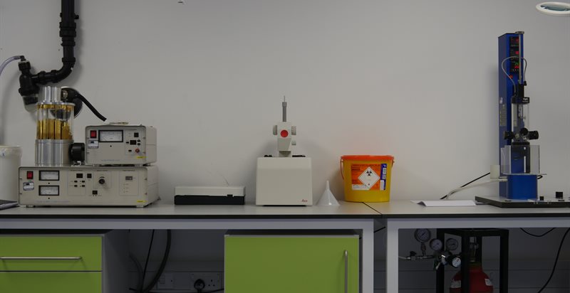
The nmRC has a fully equipped sample preparation laboratory to support its electron microscopy and chemical analysis facilities. In order for samples to be analysed by SEM, TEM, XPS and Raman Spectroscopy there are a variable number of requirements for sample pre-processing.
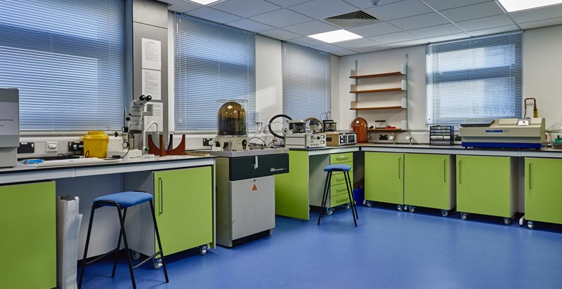
Biofluidic Microscopy (BFM) is a new integrated imaging platform that combines ultra-fast confocal imaging with the atomic force and nano-fluidic functionality. BFM enables characterisation of localised biochemical and physiological processes in three dimensions of space, time and force, thus offering a step-change capability for exploration of biological and bioinspired systems in 5 dimensions ('5D microscopy').
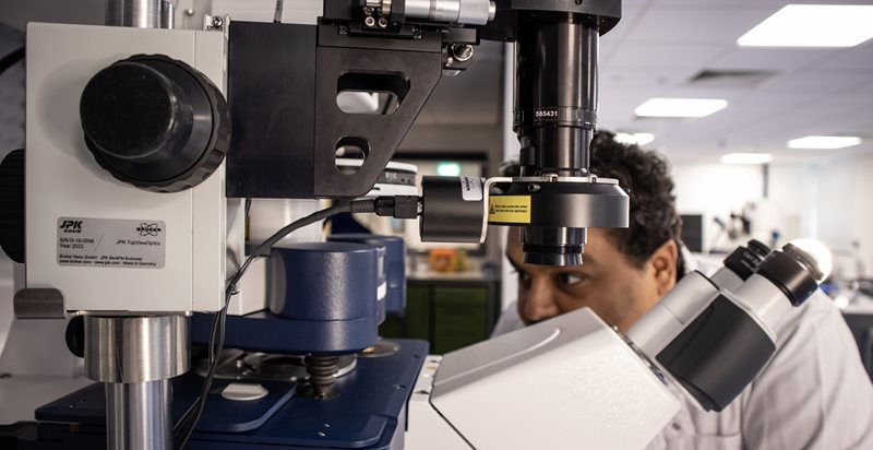
Confocal microscopy allows the visualisation of sub-cellular structures in 3D and in real-time with our incubator stage allowing live analysis of biological samples. The Airyscan 2 detector can image objects down to a resolution of 140nm. Combined with the optical tweezers we can conduct trapping and force measurement experiments simultaneously. All our confocals are equipped with correlative software so samples can be analysed across multiple instruments at the nmRC.
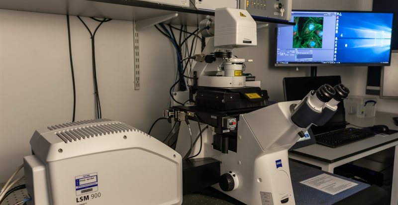
Cryo Sample Preparation
The nmRC houses a range of equipment to enable samples to be prepared under cryogenic conditions. Samples can be cryo immobilised to preserve ultrastructure and prevent damaging ice crystal formation to a depth of ~200 microns.
e-leaflets
Here you will find a selection of our e-leaflets which can be downloaded. The leaflets are a quick and easy way to find out about the range of instrumentation related to a certain technique and their key functions.
Scanning Electron Microscopy (SEM) 
Transmission Electron Microscopy (TEM) 
Atomic Force Microscopy (AFM) 
Raman Spectroscopy 
Secondary Ion Mass Spectrometry (SIMS) 
X-ray Photoelectron Spectroscopy (XPS) 
Nanofabrication Suite 
Biophysical Analysis 
Cryogenic Materials Characterisation 
Particle Sizing 