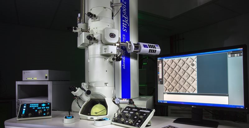
The versatile JEOL 2100+ TEM at the nmRC for nanostructural characterisation
Transmission Electron Microscopy Imaging and Analysis
TEM is an electron microscopy technique that is used to achieve spatial resolutions down to the scale of several Angstroms (~ 0.19nm). Very thin samples can be interrogated via the interaction of an electron beam as it passes through the specimen.
By monitoring what has happened to the beam of electrons in transit it is possible to both image the fine structure (by contrast) of the sample as well as describe physical and chemical characteristics.
The nmRC is home to 3 TEM instruments with state of the art cameras and detectors that offer the capacity to perform a complementary range of cutting edge analytical procedures. This ranges from bright field imaging on the nanoscale to elemental mapping, crystalline structure assessment, tomographic imaging, cryogenic imaging, dynamic reaction capture and beyond.
Get in touch with us to discuss your TEM requirements.
Key Features
- Bright field imaging
- Dark field imaging
- Electron diffraction
- Energy Dispersive X-ray Spectroscopy (EDS/EDX)
- Electron Energy Loss Spectroscopy (EELS)
- Scanning Transmission Electron Spectroscopy (STEM)
- Nanotomography (3D profiling)
- Cryogenic TEM
-
Field emission electron gun (FEG) instrument, providing a high brightness and high stability electron source for use at 100kV and 200kV
-
A point resolution of 0.19nm allows the ultrahigh resolution analysis of materials, on the nanometer scale
-
Bright Field STEM Detector
-
High Angle Annular Dark Field (HAADF) STEM Detector
-
Gatan K3 IS camera. This is a 23.6 megapixel, electron counting direct detection (DDE) camera. Capable of 150 frames per second at full view, or >3500fps at 256x256 pixels.
-
MEMS Heating holder. DENSsolutions Wildfire S3 capable of analyses up to 1300oC with millisecond heat and quench speed, and nanoscale sample drift with step changes of hundreds of degrees. Enables EELS and EDS mapping at elevated temperatures.
-
Oxford Instruments 80mm X-Max system for energy dispersive x-ray spectroscopy (EDS) analysis
-
Room Temperature Tomograpghy: Gatan 916 Room temp tomography holder with up to 80 degrees tilt. Allows tilt series acquisition to generate 3D images
-
Cryo-tomography and cryo transfer: Gatan 914 & Gatan Elsa Cryo-tomography holders including cold controller/ cryo-workstation.
-
Electrical holder. Gatan 936 DT analytical LN2 holder with temperature controller with EBIC stage option (4 electrical connections) plus Smart EBIC. Allows the electrical properties of samples to be related to the microstructure - e.g., defects in semiconductors
-
Tomography compustage for tomographic characterisation
-
Cryo-stage for low temperature observation of temperature dependent or hydrated samples
-
Ideal for low-contrast, beam-sensitive biological specimens, or other soft materials such as polymers.
Leica EM GP2 Automatic Plunge Freezer
The Leica GP2 allows cryogenic TEM imaging of hydrated biological specimens, e.g. proteins, extra-cellular vesicles etc. close to their native state. Rapidly plunging the sample into liquid ethane leads to the formation of a very thin layer of vitreous ice containing species of interest on the TEM grid, facilitating high-resolution imaging while mitigating damage from the electron beam.
-
LaB6 TEM for high throughput, high versatility analysis at 80kV or 200kV
-
Gatan OneView camera. High resolution, 16-megapixel CMOS camera for both low and high dose applications. Capable of 25 full frames per second or 300fps at 512x512 pixels.
-
Bright field STEM detector
-
Oxford Instruments X-MaxN 80 TLE EDS detector for chemical analysis
-
Gatan Enfinium EELS detector
-
HADDF detector
-
Range of specialised sample rods including heating and cryogenic stages for variable temperature work and an analytical stage for tomographic and high contrast chemical analysis
The VitroTEM Naiad facilitates fabrication of graphene liquid cells, nanosized pockets of water trapped in between two graphene sheets suitable for investigating biological and other soft matter specimens in the TEM in a native, hydrated state. The excellent mechanical and electrical properties of the graphene protect the specimen from the imaging electron beam.