3 Tesla Philips Achieva MRI Scanner
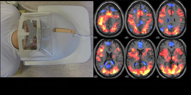
The laboratory is equipped with a 3 Tesla (3T) whole body MR system (Philips Achieva) with multi-transmit and receive capabilities. This enables the lab to offer structural and functional imaging as well as spectroscopic measurement of energy stores and metabolite levels in the brain and body.
This MRI system is equipped with a broad range of coils, for use in neurological, musculoskeletal and vascular research fields. It has capabilities to perform:
- Various fMRI stimulus devices
- Diffusion Tensor Imaging
- MR Angiography
- Perfusion
- 1H, 31P, 13C, 23Na capability
A Power Injector and physiological monitoring are also available.
Capabilities
Receive coils
We have the following (imaging) receive coils:
- 32 channel head coil (allows multi-transmit and SENSE)
- Torso XL coil (allows multi-transmit and SENSE)
- Torso coil (allows SENSE)
- 8 channel head coil (allows SENSE)
- Spine coil (allows SENSE)
- Whole body coil
- Various flex coils (circular can be wrapped around anatomy, allows SENSE)
- Birdcage head coil
- 15 channel head coil (can be used with other coils)
- 15 channel head and neck (can be used with other coils)
- 2x 16 channel Anterior coils (capable of full body imaging when simultaneously used with head coil)
- Flex extremity coils
- Dedicated MSK coils including: shoulder coil, wrist coils, knee coil and foot/ankle coil
- Microscopic coils for phalanges and other smaller samples
Spectroscopy
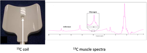
- Large and small single loop carbon coils with proton decouple coils (for 13C imaging and spectroscopy in muscle, brain and liver).
- Phosphorous coil with body coil decoupling for imaging and spectroscopy in muscle, liver and brain.
- Double flex fluorine coil with body coil decoupling (19F imaging and spectroscopy in head and body).
- Proton coils (as used for imaging).
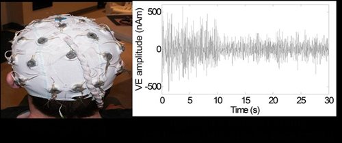
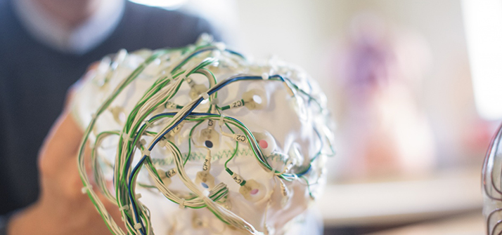
Electroencephalography (EEG)
32 or 64 channel MR compatible EEG system and caps (Brain Products GmbH). This allows functional MR and EEG signals (which measure neuronal oscillations directly) to be acquired simultaneously. Research in this area ensures advice can be given so that acquisition methods to minimise artefacts are optimal.
Electromyography (EMG)
EMG measurements can be recorded with unipolar or bipolar MR compatible amplifiers (Brain Products GmbH). This allows any muscle activity to be monitored which has a number of uses in functional imaging.
Physiological signals
Breathing, pulse rate and cardiac trace can be monitored using the Philips monitoring system.
Stimulus Presentation
The lab is equipped to deliver most forms of stimulation including: visual (using goggles or projection to a screen), auditory, somatosensory (including median nerve and vibrotactile stimulation), taste and pain (temperature induced). A separate stimulus computer is used to present paradigms to the subject and this is equipped with MATLAB, PsychoPy and Presentation (Neurobehavioural systems) for paradigm generation.
The lab also has equipment to monitor responses during a task using a fORP system. Currently available in the lab are buttons or a roller-ball but other inputs could be added.
The lab has MR compatible glass that can correct vision of +/-6 to assist people when performing tasks which require visual input.
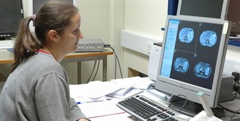
MRI scanner monitoring
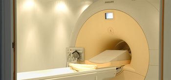
Philips Achieva 3T MRI Scanner