Cell response to surfaces
The chemical (functional groups), physical (topography) and mechanical (stiffness) properties of material surfaces play an important part in determining how cells respond to a material. Functional materials can be designed to be ‘cell instructive’, meaning the material surface is designed to produce a specific, predetermined cell response.
Cell response can be diverse; in our group we are particularly interested in controlling neuronal activity and in designing materials that control stem cell fate. The ability to affect neuronal activity will enable improved interfacing of biomedical devices with damaged neuronal tissue. Control over stem cell fate such as proliferation and differentiation is essential for the engineering of artificial tissue.
A particular area of interest in the group is the design of interactive interfaces, i.e. material surfaces that not only instruct the cell, but also respond to the cell, enabling cross talk between artificial materials and living cells.
Control of neuronal cell adhesion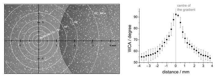
Hippocampal neurons on a radial wettability gradient (left) and profile of the wettability gradient (right). Hippocampal neurons were cultures on wettability gradients prepared by plasma deposition of hexane through a pinhole mask. The gradient was analysed by water contact angle (WCA) measurements. The number of neurons adhering to the surface decreased towards the centre of the gradient, where the surface became more hydrophobic.
Studying cell-surface interactions
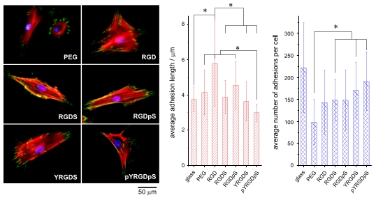
Human mesenchymal stem cell response to peptide surfaces. Left: False coloured images showing the nucleus (blue), actin (red) and focal adhesions (green). Right: quantitative analysis of the number and size of the focal adhesions. No significant difference was in the number and size of focal adhesions was observed between the phosphorylated and non-phosphorylated peptide surfaces. This suggests that cell induced dephosphorylation may render both surfaces are biologically equivalent.
Ongoing projects:
- Evaluate and control the response of neuronal stem cells to peptide surfaces.
- Study the response of stem cells to varying peptide surface densities.
- Control and measure neuronal activity through light responsive surfaces.
- Study the response of stem cells to enzyme responsive surfaces.
- Relate microparticle surface properties with stem cell adhesion and differentiation,
References
- Zelzer, M.; Alexander, M. R.; Russell, N. A., Hippocampal cell response to substrates with surface chemistry gradients. Acta Biomaterialia 2011, 7 (12), 4120-4130.
- Zelzer, M.; McNamara, L. E.; Scurr, D. J.; Alexander, M. R.; Dalby, M. J.; Ulijn, R. V., Phosphatase responsive peptide surfaces. Journal of Materials Chemistry 2012, 22 (24), 12229-12237.
- Roberts, J. N.; Sahoo, J. K.; McNamara, L. E.; Burgess, K. V.; Yang, J.; Alakpa, E. V.; Anderson, H. J.; Hay, J.; Turner, L.-A.; Yarwood, S. J.; Zelzer, M.; Oreffo, R. O. C.; Ulijn, R. V.; Dalby, M. J., Dynamic Surfaces for the Study of Mesenchymal Stem Cell Growth through Adhesion Regulation. ACS Nano 2016, 10 (7), 6667-6679.