 |
School
of Clinical Laboratory Sciences |
|
Genetics |
|
|
School
of Clinical Laboratory Sciences |
|
Genetics |
|
Constructing the tactile bacterium
A Project by Andrew Johnson and Liz Sockett
Problems faced
Many of the features of a bacterium are internal, therefore the model had to be interactive, i.e. it had to be able to be opened to explore the cells contents, and the opening mechanism needed to be simple without requiring visual co-ordination. Velcro was used to adhere the two halves of the bacterium together, which enabled a simple pull from opposite ends of the cell to open up the model. The entire model gave the students an idea of a rod shaped ‘Bacillus’ whereas the opening mechanism also allowed investigation of the cells contents.
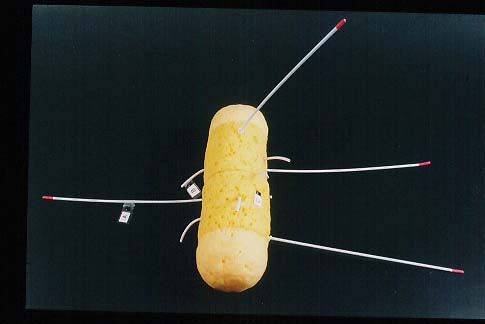
The tactile bacterium
The Solution
With respect of the contents of the cell, an ideal material for the bacterial chromosome was one length of poly-ethene rope which had one of the three coils removed. Hence creating the perfect double helix. The two ends of the rope could be fused with heat to form the circular genome. The plasmid was made in a similar fashion.
Quilt padding was chosen to represent the cytoplasm because this emulated the non solid, enveloping nature of true cytoplasm. Ideally a gelatinous material would be used but obviously this was impractical.
To represent ribosomes, ‘fimo’ modelling clay was used, as it could be moulded into the characteristic shape of 70-S ribosomes. Additionally two different coloured pieces of fimo could be used (which could be read by the colour probe) to represent the 50-S and 30-S subunits. Only a few ribosomes were included to allow the model to be kept simple and not cluttered. Tree decorating balls were used to represent the storage granules and pigment bodies due to their interesting textures.
The cytoplasmic membrane was chosen to be represented by a soft plastic cylinder. Although this material did not emulate the fluid bi-layer structure of true phospholipid membrane, and memosomes could not be built into this material, it did allow the layered structure to be more discernible when the model was pulled apart, and was durable.
Material representing the cell wall needed to echo the tough, resistant peptidoglycan of the bacterial wall which gives the cell its characteristic shape. Heavy duty cardboard tubing was chosen, which also provided a surface onto which the Velcro opening mechanism for the cell could be placed. The cell was coloured red to allow discrimination of the cell from the white of the cytoplasmic membrane (through sight or the use of the colour probe). The thickness of the material used to represent the cell wall also gave a clue to its identity.
The material to represent the glycocalyx, which a number of cells have, needed to demonstrate the gelatinous nature of this capsule. Sponge appeared to be suitable but would not be durable for continual ‘hands on’ use of the model. Therefore sheets of sponge were treated with rubber solution and stitched together to form the slime jacket for the bacterial cell model. The texture of the sponge allowed easy discrimination between the other features of the cell.
A problem with respect to flagella was that the filaments needed to be flexible and slender like true flagella. An ideal material for this was ‘curtain wire’ which is a springy coil or wire encapsulated in a plastic sheath. Pili was represented by sections of plastic sheath from electrical wire. The hollow nature of the pili were represented by this material and could possibly be detected by the students using a matchstick (or similar) to probe down the hollow core.
The overall ‘protruding layer’ structure of the model (when pulled apart) was advantageous as it allowed each layer to be recognised and labelled clearly, and also showed the origin of the flagella coming through the cell wall.
Feedback relating to the generalised bacteria model after the second visit to New College was as follows. It was suggested that the best way of labelling the model would be to have enlarge print and Braille ‘tags’ on the different features of the model which could be related to an accompanying key. Additionally a matter of safety was raised with respect of the tips of the flagella which unpredictably moved around during exploration of the model possibly catching the face of the students. To overcome this, caps were placed on the tip of each flagella which gave them a more rounded end. The cap was a sleeve of heat shrink plastic. It was during the second visit that the idea of using the more realistic plastic cylinder to represent the plasma membrane was aired. As a result of the second visit to New College to following key was created, relating to the labelled tags on the different features of the model. This was reproduced in Braille and large print, and was very brief due to the bulky nature of both large print and Braille.
Key UsedA = Flagella. Thin protein filaments used for motility.
B = Pili. Short protein protrusions, involved in attachment to surfaces.
E.g. Host body Cells
C = Capsule. Thick layer of slime, used for attachment and defence.
Pull Model Apart
D = Cell wall. Rigid structure containing peptidoglycan. Gives cell its shape.
E = Cytoplasmic membrane. Thin phospholipid bi-layer.
F = Chromosome. Single circular piece of DNA.
G = Plasmid. Small ring of extrachromosomal material.
H = Storage granule.
The small beads represent the 70-S ribosomes. The 30-S and 50-S subunits can be felt.
[Touching Science]
[ Making the bacterium ] [ Restriction Endonucleases ] [ Gene cloning models ] [ Nitrogen Cycle ]
Back to Liz Socketts home page