School of
Clinical Laboratory SciencesGenetics
Restriction endonucleases
A Project by Andrew Johnson and Liz Sockett
School of
Clinical Laboratory SciencesGenetics
Restriction endonucleases
A Project by Andrew Johnson and Liz Sockett
Restriction Enzyme Model Kit.
One reason for the construction of a restriction endonuclease kit was that it would be a good introduction to further models which were to be made depicting gene cloning experiments. An additional reason was that any models relating to restriction of endonuclease sites would also incorporate and enable revision of the basic structure of DNA.
The list of facts about restriction enzymes below, shows a summary of findings from research into the relevant amount of knowledge regarding restriction enzymes which appears to be required for A-level Biology.
Gene splicing depends on restriction enzymes (RE’s)
RE’s are naturally occurring enzymes
Specific enzymes cut at specific sights on DNA
The location of cut depends of base pairs sequence
RE’s cut both strands of DNA at slightly different points- ‘sticky ends’ are produced
Sticky end sequence depends on which one of the many RE’s are used to cut
Sticky ends allow two different pieces of DNA which have been cut by the same RE to join together under correct conditions
The base pair sequence at which the RE’s cut are unique to each RE
Common examples such as Eco R1 are useful.
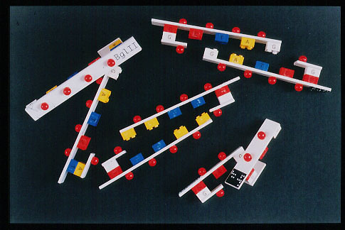
It was decided that the best way to relate all this information was through a series of interactive DNA models, which could be manipulated by the students, and be pulled apart revealing the sticky ends as though having been treated with endonuclease. Each interactive model would represent the unique sequence of DNA base pairs which would be recognised by a specific restriction enzyme.
Four models were made, showing the recognition sequence, and subsequent sticky ends produced by Eco RI, Bam HI, Bgl II, and Hpa II restriction enzymes which cut at the following sequences.
Eco R I – GAATTC Bam HI – GGATCC
CTTAAG CCTAGG
Bgl II – AGATCT Hpa II – GGCC
TCTAGA CCGG
These four restriction enzymes were chosen as examples to be made into models for a number of reasons. Eco RI is the most widely quoted and is a widely used restriction enzyme. Hpa II was chosen to demonstrate that not all restriction enzymes recognise and cut at a six base pair sequence which results in a four base pair sticky end. Bam HI and Bgl II were chosen to be represented in model form because although the whole six pair sequence which is recognised but each enzyme is different, the same four base pair sticky end is produced.
Problems faced in constructing the RE models
A major problem came to light with respect to the design of these restriction enzyme models, the solution of which dictated the relative success or failure of the model. The problem was finding a suitable material and mechanism to represent the complementary base pairs of the double stranded sections of DNA. The design and mechanism in question had a number of requirements:-
Each A,T, C and G base had to be recognisably different from each other.
Each base pair had to leave a unique complementary fit with its opposing base therefore allowing the formation of a base pair, and only allowing two base pair sticky end overhangs produced by the same restriction enzyme to fit together.
The complementary fit of corresponding bases, forming a base pair needed to be simple, not requiring visual co-ordination, but strong enough to keep both strands of DNA together until the model is pulled apart by students, simulating the action of a restriction enzyme.
The Solution
After consideration, ‘Lego’ bricks were selected to fulfil these criteria, although some modifications to each brick were undertaken to fully satisfy the above requirements.
The initial choice for the choosing the cube shaped ‘Lego’ pieces was influenced by the fact that the Lego pieces were differently coloured, allowing colour coding for each base (white for Guanine, yellow for Adenine, red for Cytosine and blue for Thymine) and that although the attachment mechanism doesn’t require visual co-ordination, it was complex enough to be modified, allowing unique binding by complementary bases, with the inhibition of attachment by any other base.
The modification of each Lego cube involved either the removal of one of two studs on the cube, or the filling in or one or two hollow stud receptors on the underneath of the cube.
Additional information which the Lego allowed to be incorporated into the model, was the number of hydrogen bonds involved in joining the base pairs together. Each Lego stud represents one hydrogen bond.
The bases were labelled with Braille on a transparent material on one side, and a printed letter, on transparent material, representing the base letter on the other side therefore allowing the colour code of the base to be seen as well as the Brailled or printed letters.
The base sequences were the major feature of these models therefore a simple backbone to each DNA strand was made from a Perspex strip. These strips represented the sugar phosphate backbone of the DNA segments and therefore to give an idea of the simple alternating ‘Sugar molecule, phosphate molecule’ nature of the backbone, red beads were glued onto the model, midway between the base pairs to represent the Phosphates. This additionally gave an indication as to exactly where in relation to the sugar and the Phosphate molecules of the backbone the restriction enzyme cuts.
Finally each model was labelled in Braille and large print. The main source of instruction and information to accompany these models was in the form of vocal instruction from a pre-recorded cassette. The following commentary was composed.
" For the following models it is assumed that the student has basic knowledge of the structure of DNA …………….. A restriction enzyme is a DNA cutting enzyme which recognises a certain sequence of A, T or G, C base pairs on a double stranded piece of DNA. These cutting enzymes are found naturally in various Prokaryotes and can be isolated and use as tools to cut DNA. There are many different restriction enzymes with different names, four models are supplied to give an example of four different codes on DNA which are recognised by different endonucleases. These enzymes are listed on an accompanying Brailled sheet along with the DNA codes which they recognise and cut at. The four enzymes are Eco RI, Bam HI, Bgl II and Hpa II. Find the model labelled Eco RI. This represents the code recognised by Eco RI endonuclease, the bases which make up the rungs of the DNA ladder are labelled on the side, either A, T, C or G. They are also colour coded. A is yellow, T is blue, G is white and C is red. The beads along the outside edges of the model represent the phosphate molecules which make up the backbone of the DNA structure. The code for Eco RI is a six letter code, wherever it occurs on DNA he enzyme will make a cut.
You will not act as the restriction enzyme, take hold of both backbones of the DNA model and gently pull them away from each other. Hopefully you haven’t broken the model and have found that the DNA has not cut straight through, but has been cut in a zig-zag fashion creating overhanging ends. These are called sticky ends in the world of biotechnology. The overhang for Eco RI is four bases which you can feel. Although these four bases are cut by Eco RI, the whole recognition sequence is six base pairs. The overhangs are called sticky ends because they will readily attach to a complementary overhang. Try this by putting the model back together.
Now explore the other two models which have a six base pair code. These models are labelled Bam HI and Bgl II. If you read the code on these models you should find that they are very similar to each other. Infact they are so similar that although recognised by different enzymes, when the DNA is cut the same sticky end overhang is produced. Observe this by pulling both models apart. The sticky end from Bam HI can join with the sticky end from Bgl II. But if you try rejoining to the Eco RI sticky end any of these others you will find that it doesn’t fit.
Whilst trying to find the ends together, notice that some bases have three studs. This represents the number of hydrogen bonds between the bases. There are two bonds between Guanine and Cytosine, whereas there are three bonds between Adenine and Thymine.
Not all endonuclease recognition codes are six bases long. This is demonstrated by the model labelled Hpa II. All these enzymes work by cutting sugar phosphate backbone of the DNA creating a nick. The DNA can be rejoined by a ligase enzyme which re-bonds the backbone. This is shown in an accompanying thermoform diagram."
[Touching Science]
[ Making the bacterium ] [ Restriction Endonucleases ] [ Gene cloning models ] [ Nitrogen Cycle ]
Back to Liz Sockett's home page