What is confocal microscopy?
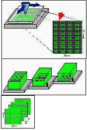
 The confocal microscope is a relatively new system which enables the optical sectioning of a 3D specimen without the need to employ some of the invasive methods used traditional histology. Information can be collected from a single focal plane of a fluorescently labelled biological specimen. A confocal pinhole excludes out-of focus light from above and below the focal plane resulting in an increase in image resolution. By moving the focal plane of the instrument step by step through the depth of the specimen a series of optical sections can be recorded. The sections collected can subsequently be reconstructed in 3D, surface rendered etc.
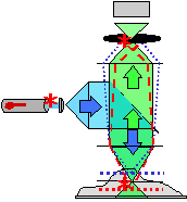
 To image the specimen point by point, a collimated, polarised laser beam is rastered stepwise in the x- and y-direction onto a dichroic mirror (beam splitter), through the objective lens of the microscope, and focused onto the specimen. The emitted, longer-wavelength fluorescent light collected by the objective lens passes through the dichroic mirror (transparent for the longer wavelength) and any necessary emission filters and then focused into a small pinhole (i.e., the confocal aperture) to eliminate all the out-of-focus light, i.e., all light coming from regions of the specimen above or below the plane of focus. Therefore, the confocal microscope does not only provide excellent resolution within the plane of section (0.25 mm in x- and y-direction), but also yields similarly good resolution between section planes (0.3 mm in z-direction). The in-focus information of each specimen point is recorded by a light-sensitive detector (i.e., a photo-multiplier) positioned behind the confocal aperture, and the analogue output signal is digitised and fed into a computer.